DISCLAIMER

The contents of this blog are solely meant for information & education purpose only.These may be the basis of actual treatment, but not necessarily. Information from other websites and journals are also included. So the author is not responsible for any inaccuracy,loss, or damage that may arise due to the use of these informations published here. I do respect copyright & always give credits to the original author(s) and thankful to them. Inspite of my utmost effort and care there can be human error. If anyone finds any violation of copyright please inform me at anesthesiatoday@gmail.com and necessary action will be taken soon as possible.
My blog is also non-commercial.


Friday, November 5, 2010

AHA CPR GUIDELINES 2010

Cardiopulmonary Resuscitation is an emergency life saving procedure consisting of  delivering effective chest compressions and effective ventilations to a victim of cardiac arrest. The American Society of Anesthesiology and European Resuscitation Council have made evidence based guidelines for the efficient and proper conduct of high quality CPR.These guidelines, being revised from time to time according to newer evidences, research and outcome help the primary care provider to offer the best care for the victims of cardiac arrest.The 2010 AHA Guidelines for CPR and ECC are based on an international evidence evaluation process that involved hundreds of international resuscitation scientists and experts who evaluated, discussed, and debated thousands of peer reviewed publications. Here is the new" guidelines(2010) in nutshell' for CPR from AHA .The major changes have been highlighted.A detailed information  of both ERC and AHA Guidelines, is available from the resuscitation council links  given below.

BASIC LIFE SUPPORT
1.Continued Emphasis on High-Quality CPR:
The 2010 AHA Guidelines for CPR and ECC once again emphasize the need for high-quality CPR, including

  • A compression rate of at least 100/min (a change from“approximately” 100/min) 
  • A compression depth of at least 2 inches (5 cm) in adults and a compression depth of at least one third of the anteroposterior diameter of the chest in infants and children(approximately 1.5 inches [4 cm] in infants and 2 inches[5 cm] in children). Note that the range of 1½ to 2 inches is no longer used for adults, and the absolute depth specified for children and infants is deeper than in previous versions the AHA Guidelines for CPR and ECC
  • Allowing for complete chest recoil after each compression
  • Minimizing interruptions in chest compressions
  • Avoiding excessive(hyper) ventilation

There has been no change in the recommendation for a compression-to-ventilation ratio of 30:2 for single rescuers of adults, children, and infants (excluding newly born infants). The 2010 AHA Guidelines for CPR and ECC continue to recommend that rescue breaths be given in approximately 1 second. Once
an advanced airway is in place,rescue breaths can  be provided at about 1 breath every 6 to 8 seconds (about 8 to 10 breaths/minute)  and need not be synchronised with chest compressions which  can be
continuous (at a rate of at least 100/min)
2.A Change From A-B-C to C-A-B
The major change made in BLS,    from airway, breathing,and circulation  the sequence has been changed to compression,airway and breathing .This is to aviod delay in delivering fast and effective chest compressions. Securing airway as the initial priority is time consuming and may not be 100% successful, especially by lone rescuers or paramedics.The vast majority of cardiac arrests occur in adults and the commonest causes for arrest are VF or pulseless VT. A witnessed cardiac arrest in these cases can be efficiently reverted with immediate defibrillation and cardiac compressions, which is life saving,  and should be the goal in BLS.. In the A-B-C sequence, chest  compressions are often delayed while the responder opens the airway to give mouth-to-mouth breaths, retrieves a barrier device,or gathers and assembles ventilation equipment.After initiating the emergency response system the next important thing is to start chest compressions.Only infant cpr is an exception to this protocol,where the previous sequence remains unchanged. That means no more looking, listening  and feeling,as this component of assessment is removed from the guidelines. In the C-A-B sequence,chest compressions will be initiated sooner and ventilation only minimally delayed until completion of the first cycle of chest compressions. It was observed that the bystanders of the arrested victims do not actively participate in CPR as they find the first step of a-b-c sequence is difficult to perform. A-B-C starts with the most difficult procedures: opening the airway and delivering rescue breaths and that is the reason why less than one third of the victims in cardiac arrest  receive by stander CPR in a witnessed cardiac arrest.. Hence  a change in sequence to C-A-B
3.Compression rate: Should be at least 100/min (rather than“approximately” 100/min). The number of chest compressions delivered per minute during CPR is an important determinant of return of spontaneous circulation (ROSC) and survival with good neurologic function
4.Compression depth: For adults has been changed from the range of 1½ to 2 inches to at least 2 inches (5 cm).(The motto is push harder and faster) Effective compressions generate critical blood flow and oxygen and energy delivery to the heart and brain.

5.Hands Only CPR: Hands Only CPR. This is technically a change from the 2005 Guidelines, but AHA
endorsed this form of CPR in 2008. The Heart Association still wants untrained lay rescuers to do Hands Only CPR on adult victims who collapse in front of them.Hands-Only (compression-only) CPR is easier for an untrained rescuer to perform and can be more readily guided by dispatchers over the telephone.It was documented that survival rates from cardic arrest of cardiac origin are same irrespective of compressions alone(hands only cpr) or  compressions with ventilations
5.Dispatcher Identification of Agonal Gasps: It is important that the rescuer  shoul be well trained to identify between  normal respirations  from agonal breaths, in order to proceed with CPR. The lay rescuer should be taught to begin CPR if the victim is “not breathing or only gasping.” The healthcare provider should be taught to begin CPR if the victim has “no breathing or no normal breathing (ie, only gasping).”This rapid breathing check should be done before activation of  emergency response system.

6.Cricoid Pressure: Routine use of cricoid pressure is not recommended as it may impede ventilation.  Studies showed that cricoid pressure can delay or prevent the placement of an advanced airway and some aspiration can still occur even with proper application.In addition, it is difficult to appropriately train rescuers in use of this maneuver.

7.Activation of Emergency Response System: Should be made after assessment of the patients' responsiveness and breathing but should not be delayed. The 2005 guidelines states immediate activation of EMS after finding an unresponsive victim.(or send someone to do so), If the healthcare provider does not feel a pulse within 10 seconds, the provider should begin CPR and use the AED when it is available.
8. Concept of team resuscitation:  For better  and  efficient delivery of  resuscitation,is emphasized.


ELECTRICAL THERAPIES INCLUDING USE OF AED AND DEFIBRILLATOR.

1.AED Use in Children Now Includes Infants
For attempted defibrillation of children 1 to 8 years of age with an AED, the rescuer should use a pediatric
dose-attenuator system if one is available. If the rescuer provides CPR to a child in cardiac arrest and does not have an AED with a pediatric dose-attenuator system, the rescuer should use a standard AED. For infants (<1 year of age), a manual defibrillator is preferred. If a manual defibrillator is not available,an AED with pediatric dose attenuation is desirable. If neither is available, an AED without a dose attenuator may be used. Automated external defibrillators with relatively high-energy doses have been used successfully in infants in cardiac arrest, with no clear adverse effects. No other major changes have bee made in electrical therapies including AED and defibrillator.

ADVANCED CARDIAC LIFE SUPPORT
1.Capnography Recommendation:  Quantitative waveform capnography is recommended for confirmation of endotracheal tube placement and for monitoring CPR quality and detecting return of spontaneous circulation based on end tidal CO2. Because blood must circulate through the lungs for CO2 to be exhaled and measured, capnography can also serve as a physiologic monitor of the effectiveness of chest compressions and to detect return of spontaneous circulation. Ineffective chest compressions (due to either patient characteristics or rescuer performance) are associated with a low Petco2 and return of spontaneous circulation is associated with an abrupt increase in ETCO2. Previously an exhaled carbon dioxide (CO2) detector or an esophageal detector device was recommended to serve this purpose.
2.Simplified ACLS Algorithm and New Algorithm:   The new circular algorithm is introduced  in 2010
The conventional ACLS Cardiac Arrest Algorithm has been simplified and streamlined to emphasize
the importance of high-quality CPR. The 2010 AHA Guidelines for CPR and ECC note that CPR is ideally guided by physiologic monitoring and includes adequate oxygenation and early defibrillation while the ACLS provider assesses and treats possible underlying causes of the arrest. There is no definitive clinical evidence that early intubation or drug therapy improves neurologically intact survival to hospital discharge.The algorithm focusses on to the basics with an increased emphasis on what is known to work: high quality CPR.


3.New Medication Protocols:

  • Atropine is not recommended for routine use in the management of PEA/asystole and has been removed from the ACLS Cardiac Arrest Algorithm.
  • The algorithm for treatment of tachycardia with pulses has been simplified. Adenosine is recommended in the initial diagnosis and treatment of stable,undifferentiated regular, monomorphic wide-complex tachycardia (this is also consistent in ACLS and PALS recommendations). It is important to note that adenosine should not be used for irregular wide-complex tachycardias because it may cause degeneration of the rhythm to VF.
4.Organized Post–Cardiac Arrest Care: 2010 (New): Post–Cardiac Arrest Care is a new section
in the 2010 AHA Guidelines for CPR and ECC. To improve survival for victims of cardiac arrest who are admitted to a hospital after ROSC, a comprehensive, structured, integrated,multidisciplinary system of post–cardiac arrest care should be implemented in a consistent manner.Treatment should include cardiopulmonary and neurologic support. Therapeutic hypothermia and percutaneous coronary interventions (PCIs) should be provided when indicated.Because seizures are common after cardiac arrest, an electroencephalogram for the diagnosis of seizures should be performed with prompt interpretation as soon as possible and should be monitored frequently or continuously in comatose patients after ROSC.
5.Initial and Later Key Objectives of Post–Cardiac Arrest Care:
1. Optimize cardiopulmonary function and vital organ perfusion after ROSC
2. Transport/transfer to an appropriate hospital or critical care unit with a comprehensive post–cardiac arrest treatment system of care
3. Identify and treat ACS and other reversible causes
4. Control temperature to optimize neurologic recovery
5. Anticipate, treat, and prevent multiple organ dysfunction.This includes avoiding excessive ventilation and hyperoxia.
6.Tapering of Inspired Oxygen Concentration:
After ROSC Based on Monitored Oxyhemoglobin Saturation,  ie, SPO2. New recommendation


ETHICAL ISSUES
The ethical issues relating to resuscitation are complex,occurring in different settings (in or out of the hospital) and among different providers (lay rescuers or healthcare personnel) and involving initiation or termination of basic and/or advanced life support. All healthcare providers should consider the ethical, legal, and cultural factors associated with providing care for individuals in need of resuscitation. Although providers play a
role in the decision-making process during resuscitation, they should be guided by science, the preferences of the individual or their surrogates, and local policy and legal requirements.
Terminating Resuscitative Efforts in Adults With Out-of-Hospital Cardiac Arrest
• Arrest not witnessed by EMS provider or first responder
• No ROSC after 3 complete rounds of CPR and AED analyses
• No AED shocks delivered
For situations when ACLS EMS personnel are present to provide care; for an adult with out-of-hospital cardiac arrest, an “ACLS termination of resuscitation” rule was established to consider
terminating resuscitative efforts before ambulance transport if all of the following criteria are met:
• Arrest not witnessed (by anyone)
• No bystander CPR provided
• No ROSC after complete ALS care in the field
• No shocks delivered
Implementation of these rules includes contacting online medical control when the criteria are met. In 2005 guidelines,no specific criteria were established

THE PEDIATRIC ADVANCED CARDIAC LIFE SUPPORT
Many key issues in the review of the PALS literature resulted in refinement of existing recommendations rather than new recommendations; 
1. Monitoring capnography/capnometry is again recommended to confirm proper endotracheal tube position and may be useful during CPR to assess and optimize the quality of chest compressions.
2.The PALS cardiac arrest algorithm was simplified to emphasize organization of care around 2-minute periods of uninterrupted CPR.
3.The initial defibrillation energy dose of 2 to 4 J/kg of either monophasic or biphasic waveform is reasonable but for ease of teaching, a dose of 2 J/kg may be used (this dose is the same as in the 2005 recommendation). For second and subsequent doses, give at least 4 J/kg. Doses higher than 4 J/kg (not to exceed 10 J/kg or the adult dose) may also be safe and effective, especially if delivered with a biphasic defibrillator.
4.On the basis of increasing evidence of potential harm from high oxygen exposure, a new recommendation has been added to titrate inspired oxygen (when appropriate equipment is available), once spontaneous circulation has been restored, to maintain an arterial oxyhemoglobin saturation ≥94% but <100% to limit the risk of hyperoxemia.
5.New sections have been added on resuscitation of infants and children with congenital heart defects,including single ventricle, palliated single ventricle, and pulmonary hypertension. The use of extracorporial membrane oxygenation , if facilities are available is stressed.
6.Several recommendations for medications have been revised. These include, not administering calcium except in very specific circumstances like hypocalcemia, calcium channel blocker overdose,
hypermagnesemia, or hyperkalemia. and limiting the use of etomidate in septic shock. Routine calcium
administration in cardiac arrest provides no benefit and may be harmful.
7.Indications for postresuscitation therapeutic hypothermia have been clarified somewhat.(see below)
8.New diagnostic considerations have been developed for sudden cardiac death of unknown etiology.
9.Providers are advised to seek expert consultation, if possible, when administering amiodarone or procainamide to hemodynamically stable patients with arrhythmias.
10.The definition of wide-complex tachycardia has been changed from >0.08 second to >0.09 second.

When a sudden, unexplained cardiac death occurs in a child or young adult, obtain a complete past medical and family history (including a history of syncopal episodes, seizures, unexplained accidents/drowning, or sudden unexpected death at <50 years of age) and review previous ECGs. All infants, children, and young adults with sudden, unexpected death should, where resources allow, have an unrestricted complete autopsy, 
preferably performed by a pathologist with training and experience in cardiovascular pathology. Tissue should be preserved for genetic analysis to determine the presence of channelopathy. It is explained as ;There is increasing evidence that some cases of sudden death in infants, children, and young adults may be associated with genetic mutations that cause cardiac ion transport defects known as channelopathies. These can cause fatal arrhythmias, and their correct diagnosis may be critically important for living relatives

NEONATAL RESUSCITATION

1.Once positive-pressure ventilation or supplementary oxygen administration is begun, assessment should consist of simultaneous evaluation of 3 clinical characteristics:heart rate, respiratory rate, and evaluation of the state of oxygenation (optimally determined by pulse oximetry rather than assessment of color)
2. Anticipation of the need to resuscitate:  during elective cesarean section
3. Ongoing assessment
4.Supplementary oxygen administration; For babies born at term, it is best to begin resuscitation with air rather than 100% oxygen.Administration of supplementary oxygen should be regulated by blending oxygen and air, and the amount to be delivered should be guided by oximetry.
5.Suctioning : There is no evidence that active babies benefit from airway suctioning, even in the presence of meconium, and there is evidence of risk associated with this suctioning. The available evidence does not support or refute the routine endotracheal suctioning of depressed infants born through meconium-stained amniotic fluid.
6.Ventilation strategies (no change from 2005)positive airway pressure may be helpful in the transitioning of the preterm baby. Use of the laryngeal mask airway should be considered if face-mask ventilation is unsuccessful and tracheal intubation is unsuccessful or not feasible.
7.Recommendations for monitoring exhaled CO2. Exhaled CO2 detectors are recommended to confirm endotracheal intubation.
8.Compression-to-ventilation ratio remains the same: The recommended compression-to-ventilation ratio remains 3:1. If the arrest is known to be of cardiac etiology, a higher ratio (15:2) should be considered.
9.Thermoregulation of the preterm infant should be considered (no change from 2005)
10.Postresuscitation therapeutic hypothermia: It is recommended that infants born at ≥36 weeksof gestation with evolving moderate to severe hypoxic-ischemic encephalopathy should be offered therapeutic hypo thermia.
11.Delayed cord clamping : There is increasing evidence of benefit of delaying cord clamping for at least 1 minute in term and preterm infants not requiring resuscitation. There is insufficient evidence to support or refute a recommendation to delay cord clamping in babies requiring resuscitation.
12.Withholding or discontinuing resuscitative efforts (Reaffirmed 2005 Recommendation): In a newly born baby with no detectable heart rate, which remains undetectable for 10 minutes, it is appropriate to consider stopping resuscitation,considering factors such as the presumed etiology of the arrest, the gestation of the baby, the presence or absence of complications, and the potential role of therapeutic hypothermia.

THERAPEUTIC HYPOTHERMIA
In adult post–cardiac arrest patients treated with therapeutic hypothermia, it is recommended that clinical neurologic signs, electrophysiologic studies, biomarkers, and imaging be performed where available, at 3 days after cardiac arrest. Currently, there is limited evidence to guide decisions regarding withdrawal of life support. The clinician should document all available prognostic testing 72 hours after cardiac arrest treated 
with therapeutic hypothermia and use best clinical judgment based on this testing to make a decision to withdraw life support when appropriate. Explained as; on the basis of the limited available evidence, potentially reliable prognosticators of poor outcome in patients treated with therapeutic hypothermia after cardiac arrest include bilateral absence of N20 peak on somatosensory evoked potential more than or equal to 24 hours after cardiac arrest and the absence of both corneal and pupillary reflexes >3 days after 
cardiac arrest. Limited available evidence also suggests that a Glasgow Coma Scale Motor Score of 2 or less at day 3 after sustained return of spontaneous circulation and presence of status epilepticus are potentially unreliable prognosticators of poor outcome in post-cardiac arrest patients treated with therapeutic hypothermia. Similarly, recovery of consciousness and cognitive functions is possible in a few post-cardiac arrest patients treated with therapeutic hypothermia despite bilateral absent or minimally present N20 responses of median nerve somatosensory evoked potentials, which suggests they may be unreliable as well. The reliability of serum biomarkers as prognostic indicators is also limited by the relatively few patients who have been studied. 


EUROPEAN RESUSCITATION COUNCIL GUIDELINES:
Reference:

Friday, October 15, 2010

EVOLUTION THROUGH REVOLUTION, COMMEMORATING ETHER DAY!

So the LORD God caused a deep sleep to fall upon the man, and he slept; then He took one of his ribs and closed up the flesh at that place.( Genesis 2:21) Who is the first Anaesthesiologist then? its Lord,The God almighty. Adam was first formed, then Eve and she was made of the man, out of his bone, and for the man (1 Co. 11:8, 9), Thus the speciality of Anaesthesiology is divine and .... Anaesthesiologists  are followers of God.! According to the law of equations, following  Anaesthesiologist is as equivalent as following God!!   .... Of course what is of more glorified act than  giving pain relief to whom who suffer from severe physical and mental agony? Even Eve was  not lucky enough to have a painless labour, a luxury  and privilege enjoyed by her descendants, made possible by this blessed branch of medical sciences. The history of anaesthesia dates back and starts from here.

Mesmer practising animal magnetism
Before the advent of anaesthesia, surgery was a terrifying prospect. Many approached surgery as if they are facing execution  and many suffered  pain, hemorrage, shock, infection  and death, due to inappropriate assessment, lack of proper agents or equipments.The pre anaesthetic era witnessed  the application of physical means  and herbal remedies for pain relief  eg:  local application of cold water, ice or pressure. Plants and extracts,Coca leaves chewed and saliva dripped into injured areas or open wounds -Inca Shamans, Alcohol with opium or hemp –“soporific sponge" (mentioned around 1200 by Nicholas of Salerno ) , Hypnosis (Its eponymous originator was Anton Mesmer (1734-1815)). etc. Amputations were performed by giving alcohol  and patients were made intoxicated  (Apuleius, a 5th century compiler) or the legs were frozen by applying ice(Refrigeration anaesthesia  by Marco AvrelioSeverino).  Egyptian surgeons apparently half-asphyxiated children undergoing circumcision by first almost strangling them. This practice sounds almost as barbarous as the operation itself.


Opium
The discovery that plant extracts, like opium (papaver somniferum), mandragora from atropa,  belladona (morning glory) Marijuana (cannabis indica) can provide pain relief, helped the primitive physicians to conduct surgeries and threw light into the pharmacological and physiological principles of pain transmission and pain management.While the physicians across the atlantic enjoyed the benefits of coca (Viennese ophthalmologist Karl Koller  used cocaine locally for ophthalmic surgeries)(1857-1944),the chinese were focussing on acupuncture


Humphry Davy
The 19th century  stood as an important mile stone  in the history of anaesthesia with the synthesis and use of atmospheric gases  and nitrous oxide by early scientific luminaries such as Black, Priestley, and Lavoisier.Of which the first gas recognised to have anaesthetic powers was nitrous oxide (N2O) which is inert, colourless, odourless and tasteless. Nitrous oxide was first isolated and identified in 1772 by the English chemist, Joseph Priestley (1733-1804). The exhilarating effects of inhaling nitrous oxide were noted by English  chemist Sir Humphry Davy (1778-1829).The term laughing gas was given to it  and N2O  was used widely for recreational purposes.It was Davy who suggested N2O for pain relief during surgery." Nitrous Air, and Its Respiration (1800)"  Davy described the different planes of  anaesthesia as stage 1: analgesia;  stage 2: delirium;  stage 3: surgical anaesthesia; stage 4: respiratory paralysis, though without appreciating the significance of each stage.. In 1824, English country doctor Henry Hill Hickman (1800-30), performed painless operations by inhalation of CO2 but was not widely recognised.
Crawford Williamson Long
Ether was first discovered by Catalan philosopher chemist Raymundus Lullius (1232-1315). Lullius called it "sweet vitriol", it was produced by distilled sulfuric acid fortified with wine and sugar.Paracelsus observed that  chickens who were given vitriol fell asleep and awaken unharmed.The first use of general anaesthesia probably dates to early nineteenth century in Japan. On 13th October 1804, a japanese physician [1]Seishu Hanaoka (1760-1835) removed a breast tumour uneventfully for which he used "Tsusensan", a herbal preparation.The first successful use of ether anaesthesia is by Crawford Williamson Long (1815-78).who may be called as the "discoverer of anaesthesia".He removed a cyst from the neck of  Mr James Venable under ether anaesthesia but this was remained  unpublished.


Horace Wells
 The anaesthetic revolution set a pace,  Horace Wells (1815-1848) demonstrated that injuring leg under N2O inhalation is painless. He himself was subjected to N2O anaesthesia for dental extraction without pain.Later on he attempted a public demonstration with N2O in january 1845 which was a failue.There was laughter and cries of "humbug".

The era of surgical anaesthesia unveils, William Thomas Green Morton (1819-1868). discovered the quality of ether that it can produce surgical anaesthesia without profound respiratory or circulatory depression. He found that a slow rate of induction allowed high margin of safety for ether. He was received an invitation to give a public demonstration at Massachussets General Hospital on friday 16 oct 1846. The patient was Edward Gilbert Abbot  and the surgeon was John Collins Warren.
William Thomas Green Morton
Morton's inhaler
The surgeon excised a vascular swelling from the neck successfully. Morton used a draw over vapouriser designed by him. on Completion of surgery  Warren announced  "Gentlemen this is no humbug"  A great discovery ,a golden mile stone in the history of anaesthesiology,the day being celebrated worldwide as "ether day".Oliver wendel homes(1809-94). suggested the name  anaesthesia for this painless state.[2]

Ether Day

Ether dome
The amphitheatre at MGH, where this demonstration took place is honoured by giving the name "ether dome"This  is now a national shrine.the  Morton's grave in Mount Auburn Cemetery near Boston bears the inscription:
WILLIAM T. G. MORTON
Inventor and Revealer of Anaesthetic Inhalation
Before Whom, in All Time, Surgery Was Agony
By Whom Pain in Surgery was Averted and Annulled
SinceWhom Science Has Control of Pain


James young simpson
In Scotland, Sir James Young Simpson (1811-1870), used Ether for the first time for relieving labour pain(1846) He was experimenting on chloroform  and proved the efficacy of chloroform by giving it to those who attended a dinner party He also tried the drug himself and published his findings in Lancet. Later he himself defended the use of chloroform for pain relief in labour as it was against religion and belief. Chloroform use also had a short history. It is a colourless volatile liquid with a characteristic smell and a sweet taste.It was discovered in1831 by American physician Samuel Guthrie (1782-1848);

John snow
In England, at least, the practice of anaesthesia during childbirth won greater respectability following its widely-publicised use on Queen Victoria. The delivery in 1853 of Victoria's eighth child and youngest son, Prince Leopold, was successful: chloroform was administered by Dr John Snow (1813-1858) of Edinburgh, the world's first anaesthesiologist/anaesthetist. He used chloroform in a hand kerchief for inhalation and Queen victoria never had pain during child birth.The contributions of John snow include
  • Description of stages of anaesthesia based on patient's responsiveness
  • Developed ether inhalers,  and an ether vapouriser which is thermocompensated.
  • Development of agent specific chloroform vapouriser
  • Described the minimum anaesthetic concentration to prevent movement  which led to the future discovery of  MAC
  • Published books  and journals on chloroform and ether
  • Joseph clover
  • Conducted epidemiological surveys  and proved that cholera is transmitted by water.
 Dr Joseph Clover (1825-1882) developed  the first apparatus to provide chloroform in controlled concentrations; in 1862 and a "portable regulating ether-inhaler" in 1877. This was to minimise the complications  associated  with the use of chloroform

The other Inhalation Anaesthesia landmarks include
    Boyles apparatus
  • Introduction of first anaesthesia machine in 1917 by Henry Boyle
  • CO2 absorption canister in 1924 by Rudolf Waters
  • Endotracheal intubation in 1920 by  Ivan Magill.
  • Introducton of halothane in 1950 


    Epoch of Regional Anaesthesia[3]
    • Karl Koller( 1857-1944)  an ophthalmologist from vienna introduced cocaine for topical ophthalmic anaesthesia
    • William Halstead, described nerve blocks of face and arm 1886,
    • Description of spinal anaesthesia by Leonard Corning. He coined the term spinal anaesthesia. He used cocaine into the spinal canal
    • Regional Anaesthesia
    • August Bier, further research  on  spinal anesthesia in 1898.
    • Virginia Apgar promotes Regional analgesia in childbirth 1930.
    • Arthur Barker hyperbaric spinal anaesthesia by addition of glucose  1935
    • August Bier, intravenous regional anaesthesia in 1908
    • Martinez Curbelo from cuba  continuous epidural anaesthesia  1949
    Intravenous Anaesthesia
    • 1903 Barbitone synthesis by Fischer, Berlin, Nobel Prize.
    • 1932 Evipan used IV by Weese Schraff, & Rheinoff.
    • 1934 Lundy, Mayo Clinic, used Sodium Pentothal.
    • 1942 Curare first used clinically by Griffith and Enid johnson in Montreal
    • 1857 Claude Bernard published that curare blocked the NMJ.
    • 1940 Bennett used curare to modify seizures induced by metrozol.
    Modern anaesthesia
    • Starts from 1980
    • Standards for Basic Anesthetic Monitoring ASA, House of Delegates 10/21/1986.
    • Anesthesia Patient Safety Foundation 1984.
    • International anaesthesia research society 1922 by mcMechaan
    • BJA 1923
    • Association of anaesthetists of great britain and ireland 1932. 

    The modern Anaesthesiologist is a physician  and primary care provider who is concerned with the total wellbeing of his patients. Other than the safety and  comfort of patient in operation theatre, the anaesthesiologist's services extend outside the operating room as well. They are routinely asked to monitor or sedate patients for lithotripsy, MRI, cardiac catheterisation, electroconvulsive therapy, fluroscopy etc. They are considered to be the pioneers in cardiopulmonary resuscitation.  The anaesthesiologist is actively involved in  Intensive care, Trauma care, Pain management and complex specialised surgical procedures

    Reference:
    1). Matsuki A: Seishu Hanaoka, Japanese pioneer in anesthesia. Anesthesiol1970; 32:446-50 Fenster J M: 
    2). Sykes: Essays on the First Hundred Years of Anaesthesia. Robert E. Kreiger Publishing Co. 1972.
         Wolfe & Menczer: I Awaken to Glory. Boston Medical Library, 1994.
    3)  An article on  Anesthesiology yesterday, today and tomorrow by Mr.Adolph H. Giesecke MD.UTX Southwestern Medical Center, Dallas T
    4)  Wylie and Churchil Davidson  Practise of Anesthesia.   7th edition.; Pp 01-15
    5)  Images :
         http://en.wikipedia.org/wiki/Ether_Dome
         www.general-anaesthesia.com/images/ether-inhaler.html
         http://www.bmj.com/content/2/4475/536.full.pdf+html

    Wednesday, September 22, 2010

    THE ROLE OF CLASSIC LMA IN DIFFICULT AIRWAY.

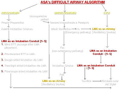
    The recent development in the management of "difficult airway" is the incorporation of LMA into its algorithm. Since the introduction of LMA in 1981 they have been widely evaluated for their efficacy in managing difficult airways and have been proven effective in CICV situations.According to the 'Practice Guidelines for Management of the Difficult Airway' by the ASA task force, the use of supraglottic airway devices including LMA, Combitube or other suitable supraglottic airway as rescue devices in the “cannot intubate cannot ventilate” situation, is highly recommended. The advice switches from an earlier emphasis on laryngoscopy and intubation to an emphasis focusing on ventilation and oxygenation.The present topic concentrates only on the algorithm where LMA can be used as an intubation conduit. Eventhough intubating LMA is widely used to manage difficult airway, classic LMA alone is included in the algorithm because of its simplicity and safety in inexperienced hands. Watch the following flow charts, photographs, and a video which are self explanatory.
    Classic LMA

    • Developed by Dr. Archie I J Brain, a British anesthetist in 1981.
    • First successful use of prototype LMA in failed intubation situation in a 114 kg patient in February 1983.
    • Dr. Archie Brain publishes his first series of 23 patients undergoing routine airway management by the LMA in 1983.
    • 21 cases of difficult intubations were managed by the LMA and reported in literature between 1983 - 1987.LMAs are commercially available since 1987.
    • How to insert:
      The standard technique:
      Why and how LMA has found a unique place in Algorithm?
    • Simple to use
    • Non-invasive design as it does not invade trachea
    • An easy to learn & easy to teach quality.
    • Improved success rate with minimal tissue trauma in first attempt
    • Ability to be used as a ventilatory device as well as an aid to intubation.
    • Immediate airway access compared to invasive techniques
    • Less gastric inflation and subsequent regurgitation.
    Alternate methods for placement of LMA:

    • Insert the deflated LMA with the laryngeal aperture facing cephalad and rotate it 180º as the cup enters the pharynx, Specially useful in patients with restricted mouth opening or hindrance to placement by tongue.
    • Insert the LMA from the side of the mouth in emergency for example from bedside using thumb
    • Insert the LMA with the cuff partially inflated when the deflated cuff gets folded.
    • Insert the LMA with the aid of a laryngoscope again in difficult insertion due to large tongue, edentulous airway or due to restricted mouth opening.
    • Insert LMA with the aid of stylet or a bougie as guide.
    • In patients with a restricted mouth opening, LMA can be placed retro molar and subsequently the LMA tube is brought forward to lie centrally
    The Algorithm:  Click on the image to enlarge.


                    ALGORITHM EXPLAINING THE ROLE OF LMA (EXPANDED)
                                   Click on the image to enlarge


    LMA AS AN INTUBATION CONDUIT:

    • Blind passage of ETT through LMA: After placement of LMA by any one of the above techniques, the lubricated endotracheal tube is passed blindly through LMA and placement confirmed by auscultation, chest movement or by capnogram. The following table helps you to choose the correct size of ETT to be passed.
    • In an attempt to insert the LMA blindly one may encounter 2 levels of obstruction within the LMA.The first one is at the level of the aperture bar and the second one is at the level of glottic opening.              
                                                                                                                                                                                                                                                                                                                                                             
      To overcome the first level of obstruction, the ETT is slightly bend and turned to one side while passing.  The second level of obstruction is overcome by flexing the neck while insertion

    • Premounted ETT in LMA with insertion:To avoid the problems associated with obstruction, the ETT is premounted on LMA and inserted. Subsequently manipulated to enter the glottis.
    • Bougie aided intubation through LMA: The LMA is inserted cuff inflated and placement confirmed. A bougie is passed through the ETT and manipulated into the larynx. The LMA is deflated and removed  and a lubricated ETT is passed over the bougie into the trachea

    • Trachlight aided intubation via LMA:  After placement of LMA a trachlight (lighted wand) is used to identify the laryngeal inlet and ETT is passed over it.
    • Fiberoscope aided intubation via LMA: Similar to trach light, a fiberoptic bronchoscope is passed into the LMA to reach the trachea and subsequent intubation over bronchoscope is done.
    Video showing blind intubation over LMA


     When no aids are available in CVCI situation the algorithm advises on cricothyrotomy and transtracheal jet ventilation.An anteriorly placed larynx can be managed, in the absence of  stylet or bougie by simple manipulation of the ETT, by anchoring it on middle finger to bring forward the tip of ETT.

    The video and a few photographs are reproduced with permission from Dr.Rashid M Khan and Dr.Naresh Kaul of Khoula hospital Muscat, who conducted a workshop on "difficult airway" at ibri hospital in March 2010. The author is much thankful to them.
    Ref:1)ASA, Practice guidelines for the management of difficult airway -2003 .   www.asahq.org/publicationsAndServices/Difficult%20Airway.pdf
           2)Leader GL, facilitation of insertion of Laryngeal mask, Anaesthesia 1991;46:987                     
           3)The Difficult Airway in Adult Critical Care.: Supraglottic Airway ,www.medscape.com

    Tuesday, August 31, 2010

    ANAESTHESIA FOR STRABISMUS SURGERY.

    One of the commonly performed ophthalmic surgery in the pediatric age group.The surgical procedure is simple and short but often associated with unexpected peri operative complications, but most of them can be successfully managed by immediate intervention by a vigilant anaesthetist.The anaesthetic concerns include controversial use of suxamethonium for induction, difficult airway, high incidence of post operative nausea and vomiting, systemic effect of topical medications,other associated congenital mal formations, oculocardiac reflex, need for post op analgesia and propensity for malignant hyperpyrexia.

    Suxamethonium:  Suxamethonium if used for induction can cause sustained contracture of intraocular muscles and this may affect the forced duction test performed by surgeons during surgery to estimate the amount of restriction in movement of extraocular muscles. Hence to ensure patient immobility during surgery non depolarising agents like atracurium, vecuronium or rocuronium are considered. But because of its faster action,effective relaxation of laryngeal muscles and that raise in intra ocular pressure is not very significant in strabismus surgery,some anaesthetists still prefer[1] suxamethonium for induction.The use of suxamethonium also helps the surgeon, as the globe adducts exposing a large bare area of sclera allowing for easy re insertion of cut lateral rectus[2]
    Incidence of Difficult Airway: Patients with squint may have associated congenital malformations like Down's syndrome,Marfans syndrome or Muscular dystrophies, with involvement of airway. Careful assessmemt of airway is mandatory before planning anaesthetic management.
    Effect of medications placed on eye:  Eye drops are readily absorbed through hyperemic, incised conjunctiva causing systemic effects Phenylephrine is placed in the eye to produce mydriasis and haemostasis, however absorption of the phenylephrine can cause profound systemic vasoconstriction and hypertension.It can also cause arrhythmia and head ache. To prevent systemic hypertension only 1 to 2% phenylephrine should be used and only one drop should be put into each eye.Adrenaline(2%) cause hypertension & arrhythmias Timolol (B-blocker) causes bradycardia, hypotension & exacerbation of asthma  Phospoline iodide(echothiophate iodide) is a long acting anti-cholinesterase used in glaucoma prolongs suxamethonium induced muscle relaxation.A patient who has been treated with echothiophate iodide can retain low blood levels of pseudocholinesterase for weeks or even months after discontinuing the medicine.Thus the use of suxamethonium is contraindicated when phospholine iodide is used for fear of post op apnea.[3]systemic effects of cyclopentolate hydrochloride include disorientation dysarthria and seizures.
    Oculocardiac reflex: Bernard Ashner and Guiseppe Dagnini first described this reflex in 1908.This reflex is a trigemino vagal reflex and is triggered by pressure on the globe or by traction on the extraocular muscles. The reflex is also triggered by ocular trauma, retrobulbar block, severe pain or by orbital compression due to hematoma or edema. the afferent impulse travels via the long and short ciliary nerves to the ciliary ganglion, then continues to the gasserian ganglion along the ophthalmological division of the trigeminal nerve and terminates at the main trigeminal sensory nucleus in the floor of the fourth ventricle. The efferent impulse travels by way of of the nucleus of the vagus nerve to the vagal cardiac depressor nerve, producing negative inotropic and conduction effects consisting of bradycardia, nodal rhythm, ectopic beats, ventricular fibrillation and rarely asystole.The surgeon should be informed immediately when this arrhythmia develops, to remove pressure or traction on the globe.

    The treatment of oculocardiac reflex should be immediate and with intravenous injection of atropine at doses of 0.005 to 0.4 mg/kg. Anticholinergic premedication helps to prevent occurence of this reflex.Recurrent episodes require local infiltration of lignocaine near the extrinsic muscles.IV Epinephrine 6-12 mg  is used for patients with cardiovascular collapse. IV Lidocaine 1.5-2 mg/kg is given for Ventricular arrhythmias along with cardiac massage. Retrobulbar block with 1 to 3 ml of 1% or 2% lidocaine (Xylocaine) may prevent oculo cardiac reflex by blocking the afferent limb of trigeminal vagal reflex.
    Malignant hyperpyrexia: Patients with strabismus have a higher incidence of malignant hyperthermia.In cases where susceptibility to malignant hyperpyrexia or a family history is suspected pre-treatment with dantrolene is required[3]. Study of phenylketopyruvate serum levels can be useful in predicting susceptibility to malignant hyperthermia in a patient with a questionable family history[3].The triggering agents include suxamethonium and halothane and hence the preferred general anesthetic regimen for patients with  susceptibility to malignant hyperthermia is propofol,fentanyl, nitrous oxide, and a non depolarising muscle relaxant.Classicaly malignant hyperthermia occurs intraoperatively and results in rapid rise in temperature, muscle rigidity, dysrrythmias, rhabdomyolysis, acidosis and hyperkalemia. Approximately, one half of patients who develop muscle rigidity after succinylcholine are susceptible to malignant hyperthermia by the muscle biopsy and contracture test. In these patients, if creatinine phosphokinase level is more than 20,000 IU, malignant hyperthermia susceptiblity is strongly suggested. If massester muscle spasm occurs, a muscle biopsy and contacture test is indicated to confirm malignant hyperthermia.[4].The treatment of malignant hyperpyrexia include discontinuation of all anaesthetic agents, 100% oxygen,dantrolene 2.5mg/kg IV,rapid cooling to bring down body temperature correction of acidosis and hyperkalemia, and ventilatory support.
    Post operative nausea and vomiting:is very common following strabismus correction. The exact mechanism is not known. It may be secondary to altered visual perception or an oculoemetic reflex, which is analogous to the oculocardiac reflex.It is more common in opioid premedicated patients.Oral midazolam 0.5 mg/kg seems to be a better premedicant for strbismus cases.Intraoperative use of metoclopramide 0.1-0.15mg/kg IV,droperidol 70 mic/kg,ondansetron 0.1mg/kg, and intravenous induction of anaesthesia by propofol etc, helps to reduce the incidence of PONV.[5]
    Post operative pain management:is also equally important to reduce pain and discomfort in children.rectal paracetamol or diclofenac suppositories are commonly used for this purpose.Pre operative subtenon's instillation of levobupivacaine is also helpful.
    Anaesthetic management:  Strabismus surgery in adult can be performed under local anaesthesia(retrobulbar or peribulbar block) with or without sedation. Adult Un-coperative patient can be managed with total intravenous anaesthetic technique with sedative and narcotic drugs. Children will always require general anaesthesia for corrective surgery. Premedication may be given with oral midazolam 0.5 mg/kg along with atropine 0.02 mg/kg. Inhalational induction with sevoflurane in oxygen and nitrous oxide,fentanyl 1mg/kg IV,rocuronium 1 mg/kg IV or atrcurium 0.5 mg/kg IV, proseal LMA/ETT, controlled ventilation.Intravenous induction is with fentanyl 1mg/kg, propofol 2.5mg/kg,vecuronium/atracurium with nitrous oxide in oxygen and isoflurane.The use of neuromuscular monitoring is strongly advised and ECG monitoring is mandatory. It is essential to maintain normocarbia throughout the procedure.Extubation or removal of LMA attempted in deep plane of anaesthesia.Intraop prophylaxis for PONV with ondansetron or metoclopramide in suggested doses should be administered.
    References:
    1)J.C Stanley, Hand Book of Clinical anaesthesia, Chrchill livingstone, 1996.
    2)D Abrams, British Journal of Ophthalmology,1984,64:218
    3)Eugene M. Helveston, M.D.Surgical Management of Strabismus: A practical and updated approach, 5th edition;http://telemedicine.orbis.org/bins/content_page.asp?cid=1-2161
    4)Practical case notes;Dr. R.C. Agarwal,Dept. of Anaesthesiology & Critical Care, Bhopal Memorial Hospital & Research Centre, Raisen by-pass Road Karond, Bhopal.
    5)Kenneth Davison, Clinical Anaesthesia Procedures of MGH, fifth edition.
    Image cortesy: www.nysora.com(subspeciality/ophthal)

    Thursday, July 15, 2010

    WIRELESS PAC CLINIC, THE SCOPE!

    Do you think the PAC clinics have started disappearing? Are they essential for pre anaesthetic evaluation? This issue has recently been emerged as a topic for discussion in some Anaesthesia conference. Eventhough at first thought,  you may feel this is an absurd statement,those who support this notion give some valid reasons. They strongly argue that ASA 1 and 2 patients are not benefitted from PAC as they are in good health or in mild systemic illness which is well controlled.These patients can be directly admitted to the ward by surgeons or they can report as day care cases, which will reduce the work strain of staff and doctors and  that this is cost effective.This method also helps to reduce the waiting time of patients in PAC clinic. For ASA 3 and 4 pre anaesthetic check up and follow up are done in ward  on a multidisciplinary approach.

    The author somehow feels this is inappropriate. A well conducted PAC clinic is essential for the proper assessment of all types of patients including ASA 1 and 2. Now office based  and day care surgeries are commonplace everywhere and patients seen in PAC clinic can be sent to office or day care suite direct,on the day of surgery without getting them admitted to the hospital.Even in a properly conducted preanaesthetic check up, the clinician missed certain vital informations in ASA 1 and 2 cases, which led to serious intra operative complications.Thus the  necessity of a thorough and proper evaluation aided by available investigations is stressed.. Look into some of the following clinical scenario.

    Wednesday, June 30, 2010

    MISDIRECTED CENTRAL VENOUS CATHETER

    Scenario 1. A 23 yr old patient with antepartum hemorrhage was taken to OT for Caesarean section. She had profuse bleeding on table  but could be controlled following delivery of baby, with blood transfusion and colloids. The chest on auscultation revealed fine basal creps and she had persistent hypotension.A central venous catheter was introduced through the right internal jugular vein. Confirmation of placement done by aspiration for free flow and rapid infusion of fluids.But the free flow was not obseved at a catheter length of 14 cm or13 cm but free flow observed at 11 cm.The patient remained ventilated and was shifted to the icu and CXR was  taken The tip of the catheter appeared to be in the right subclavian vein  with a slight angulation. A repeat procedure was deferred as the patient had coagulopathy and that the clinical confirmatory tests are satisfactory. The next day afternoon patient was complaining of raspiratory distress with tachypnea and pain on right side of chest. Auscultation revealed absent breath sounds on right side  with dullness on percussion. A repeat CXR showed  displacement of catheter tip, with massive pleural effusion on the right side which was drained subsequently.

    Monday, June 21, 2010

    ARTIFICIAL BLOOD, HOPE FOR THE FUTURE?

    A sigh of relief, the tension is relieved as the gynaecologist took the baby out with great  difficulty through the incision. The  indication for caesarean section was severe fetal distress and the OT atmosphere has now turned pleasant as the paediatrician reported that the APGAR score is 9. The anaesthesiologist is busy preparing  oxytocin infusion and sedation.The patient who was given regional anaesthesia is now anxious to see her baby.But the enjoyment did not last long as the surgical team noted profuse bleeding with a flabby uterus not responding to ergometrine or oxytocin.A request for issuing blood was  sent to blood bank but the request was returned ,stating that the husband refused any kind of blood transfusion as he is a strict Jehovah's witness which he didn't mention at the time of taking the consent!

     Jehovah's witnesses, a fellowship of more than 1 million americans, object to the administration of blood in any form for any indication.This objection is based on "THE HOLY BIBLE"  Acts 15:28,29 which states "For it has seemed good to the Holy Spirit and to us to lay on you no greater burden than these requirements: that you abstain from what has been sacrificed to idols, and from blood, and from what has been strangled, and from sexual immorality. If you keep yourselves from these, you will do well. Farewell.” According to them blood removed from the body should be discarded "You should pour it upon the ground as water"  (Deuteronomy 12:24.)This makes pre operative blood conservation technique like preoperative phlebotomy and storage impractical
    The search for an oxygen carrying blood substitute started with world war II as the military realized the difficulties of whole blood transport and storage.Subsequently several trials were made to bring out an ideal blood substitute using chemicals which can carry oxygen.
    1)Perfluorocarbon compounds:Of the various substances that carry or facilitate the transport of oxygen the perfluorocarbons gave the most promising results.They were created by replacing H2 atoms of hydrocarbons with flurine. This was following the first real success in  "fluid breathing system" proposed by Leland Clark in 1966. He found that oxygen and carbondioxide are very much soluble in fluorocarbon liquids and can be used for artificial ventilation of the lungs which are immersed in these compounds where satisfactory oxygen uptake  and giving out of CO2 takes place by the alveoli through the liquid media, in the absence of external oxygen supply.He performed his experiment in anaesthetised rat where the animal is paralysed intubated and immersed in PF liquid.After bubbling oxygen through the liquid this is pumped into the animal's lungs and recirculated. Most of the animals kept in the fluid for upto an hour survived for several weaks. Subsequently trials were made in humans during war time.               Figure: Demonstrating a living mouse immersed  in  perfluorocarbon compound along with a goldfish.
    • Fluosol-DA was the most notable compound among PFCs, regarded as a first generation PFC.It contained perfluorodecalin and perfluorotripropylamine emulsified with Pluronic F-68  It was approved by the FDA for use in percutaneous transluminal coronary angioplasty initially but was subsequently withdrawn(see below)
    • They have high affinity for oxygen approximately 10-20 times greater than plasma.The  oxygen content of PFCs  is directly proportional to oxygen partial pressure and are most efficient as oxygen carriers at a partial pressure of more than 300mmHg, which limited their use, as patients needed high inspired oxygen concentration. A short intravascular half life, unstable at extreme temperatures, Low oxygen carrying capacity ,poor shelf life and adverse effects such as acute complement activation and disruption of pulmonary surfactant, all lead to their withdrawal  from the market. But still these compounds have found some place in "Liquid ventilation" of lungs in ARDS.

    Friday, May 14, 2010

    HOW TO WEAN AND WHEN?

    A patient is considered fit for weaning from ventilator when he is conscious responsive and hemodynamically stable and when the pathology which necessitated mechanical ventilation has been resolved adequately.Improved patient outcomes and decreased costs are two benefits of implementing a protocol for early weaning from ventilator.A simplified approach to weaning off ventilator is described below.

    1).Criteria to determine whether a patient can be given a trial of spontaneous breathing.
    Repiratory criteria:
    • PaO2 > 60 mmHg on Fio2 40-50% and peep less than 5-8 cmH2O
    • PaCO2 normal or baseline
    • Adequate inspiratory effort
    Cardiovascular criteria:
    • No evidence of myocardial ischemia
    • Heart rate less than 140/mt.
    • Blood pressure normal without inotrops or minimal inotropic support eg: dopamine <5 mic/kg/mt
    Adequate mental status:
    • GCS >13 and arousable
    Absence of correctable comorbid conditions:
    • Afebrile
    • No significant electrolyte abnormalities
    2) Criteria to determine whether patients can tolerate spontaneous breathing trial
    • Tidal volume               5-7 ml/kg                  threshold    >4-6 ml/kg
    • Respiratory rate          10-18beats/mt          threshold    <30/mt.
    • RR/VT ratio                20-40 /LTR                threshold 100/LTR
    • Max insp. pressure     -90to -120 cm H2O     threshold  -15 to -30 cm H2O
    The RR/VT ratio or rapid shallow breathing index is a useul predictor. Value above 105/LT, 95% of the attempts are successfull.

    Friday, May 7, 2010

    STRESS AND ANAESTHETIST

    The previous night duty was so busy and i was tired out.The exploratory laparotomy went upto 2 am in the morning.Today i got up late and found my bus just leaving.My colleague staying next door offered me a lift but i reached hospital late.The morning ICU rounds already started and i tried to hide my self behind the team members, in order to escape notice of the consultant.The rounds finished and it was my turn to present the previous day's cases.It didnt go smooth as  I was drowsy  and was not able to concentrate. The consultant asked me to attend the elective  cases  in surgery OT where the list was also heavy with 3 major cases. While preparing for the cases, the  hospital clerk handed over  the university  notification for the venue and timings for final masters exam, scheduled  next month.The second case in my OT had a stormy recovery with laryngospam and agitation which was so difficult to control, and finally i am totally upset when the school principal telephoned and conveyed the message that my daughter is sick and will be brought to OPD.

    Stress can be defined as mental, emotional or physical strain or tension which is an integral part of everyone's life.Even though moderate stress is necessary for the optimal function of human beings undue stress may have physiological and psychological impact(1) Stress occurs when there is a perceived imbalance between the demands being made and the ability to meet those demands.It is a pattern of strain produced by excessive urgency or pressure.  According to Cooper  stress is negatively perceived quality which could cause physical and mental ill health.

    Most of the doctors are found to have Type A personality featuring insecurity of status and a high amount of anxiety. This personality type is often associated with increased aggression and a constant sense of time urgency and mental tension.They are more prone for stress related responses.They also tend to have a higher incidence of coronary artery disease and may have problems coping with and responding to difficult situations. This may lead on to psychiatric problems in the future.