One of the commonly performed ophthalmic surgery in the pediatric age group.The surgical procedure is simple and short but often associated with unexpected peri operative complications, but most of them can be successfully managed by immediate intervention by a vigilant anaesthetist.The anaesthetic concerns include controversial use of suxamethonium for induction, difficult airway, high incidence of post operative nausea and vomiting, systemic effect of topical medications,other associated congenital mal formations, oculocardiac reflex, need for post op analgesia and propensity for malignant hyperpyrexia.
Suxamethonium: Suxamethonium if used for induction can cause sustained contracture of intraocular muscles and this may affect the forced duction test performed by surgeons during surgery to estimate the amount of restriction in movement of extraocular muscles. Hence to ensure patient immobility during surgery non depolarising agents like atracurium, vecuronium or rocuronium are considered. But because of its faster action,effective relaxation of laryngeal muscles and that raise in intra ocular pressure is not very significant in strabismus surgery,some anaesthetists still prefer[1] suxamethonium for induction.The use of suxamethonium also helps the surgeon, as the globe adducts exposing a large bare area of sclera allowing for easy re insertion of cut lateral rectus[2]
Incidence of Difficult Airway: Patients with squint may have associated congenital malformations like Down's syndrome,Marfans syndrome or Muscular dystrophies, with involvement of airway. Careful assessmemt of airway is mandatory before planning anaesthetic management.
Effect of medications placed on eye: Eye drops are readily absorbed through hyperemic, incised conjunctiva causing systemic effects Phenylephrine is placed in the eye to produce mydriasis and haemostasis, however absorption of the phenylephrine can cause profound systemic vasoconstriction and hypertension.It can also cause arrhythmia and head ache. To prevent systemic hypertension only 1 to 2% phenylephrine should be used and only one drop should be put into each eye.Adrenaline(2%) cause hypertension & arrhythmias Timolol (B-blocker) causes bradycardia, hypotension & exacerbation of asthma Phospoline iodide(echothiophate iodide) is a long acting anti-cholinesterase used in glaucoma prolongs suxamethonium induced muscle relaxation.A patient who has been treated with echothiophate iodide can retain low blood levels of pseudocholinesterase for weeks or even months after discontinuing the medicine.Thus the use of suxamethonium is contraindicated when phospholine iodide is used for fear of post op apnea.[3]systemic effects of cyclopentolate hydrochloride include disorientation dysarthria and seizures.
Oculocardiac reflex: Bernard Ashner and Guiseppe Dagnini first described this reflex in 1908.This reflex is a trigemino vagal reflex and is triggered by pressure on the globe or by traction on the extraocular muscles. The reflex is also triggered by ocular trauma, retrobulbar block, severe pain or by orbital compression due to hematoma or edema. the afferent impulse travels via the long and short ciliary nerves to the ciliary ganglion, then continues to the gasserian ganglion along the ophthalmological division of the trigeminal nerve and terminates at the main trigeminal sensory nucleus in the floor of the fourth ventricle. The efferent impulse travels by way of of the nucleus of the vagus nerve to the vagal cardiac depressor nerve, producing negative inotropic and conduction effects consisting of bradycardia, nodal rhythm, ectopic beats, ventricular fibrillation and rarely asystole.The surgeon should be informed immediately when this arrhythmia develops, to remove pressure or traction on the globe.
The treatment of oculocardiac reflex should be immediate and with intravenous injection of atropine at doses of 0.005 to 0.4 mg/kg. Anticholinergic premedication helps to prevent occurence of this reflex.Recurrent episodes require local infiltration of lignocaine near the extrinsic muscles.IV Epinephrine 6-12 mg is used for patients with cardiovascular collapse. IV Lidocaine 1.5-2 mg/kg is given for Ventricular arrhythmias along with cardiac massage. Retrobulbar block with 1 to 3 ml of 1% or 2% lidocaine (Xylocaine) may prevent oculo cardiac reflex by blocking the afferent limb of trigeminal vagal reflex.
Malignant hyperpyrexia: Patients with strabismus have a higher incidence of malignant hyperthermia.In cases where susceptibility to malignant hyperpyrexia or a family history is suspected pre-treatment with dantrolene is required[3]. Study of phenylketopyruvate serum levels can be useful in predicting susceptibility to malignant hyperthermia in a patient with a questionable family history[3].The triggering agents include suxamethonium and halothane and hence the preferred general anesthetic regimen for patients with susceptibility to malignant hyperthermia is propofol,fentanyl, nitrous oxide, and a non depolarising muscle relaxant.Classicaly malignant hyperthermia occurs intraoperatively and results in rapid rise in temperature, muscle rigidity, dysrrythmias, rhabdomyolysis, acidosis and hyperkalemia. Approximately, one half of patients who develop muscle rigidity after succinylcholine are susceptible to malignant hyperthermia by the muscle biopsy and contracture test. In these patients, if creatinine phosphokinase level is more than 20,000 IU, malignant hyperthermia susceptiblity is strongly suggested. If massester muscle spasm occurs, a muscle biopsy and contacture test is indicated to confirm malignant hyperthermia.[4].The treatment of malignant hyperpyrexia include discontinuation of all anaesthetic agents, 100% oxygen,dantrolene 2.5mg/kg IV,rapid cooling to bring down body temperature correction of acidosis and hyperkalemia, and ventilatory support.
Post operative nausea and vomiting:is very common following strabismus correction. The exact mechanism is not known. It may be secondary to altered visual perception or an oculoemetic reflex, which is analogous to the oculocardiac reflex.It is more common in opioid premedicated patients.Oral midazolam 0.5 mg/kg seems to be a better premedicant for strbismus cases.Intraoperative use of metoclopramide 0.1-0.15mg/kg IV,droperidol 70 mic/kg,ondansetron 0.1mg/kg, and intravenous induction of anaesthesia by propofol etc, helps to reduce the incidence of PONV.[5]
Post operative pain management:is also equally important to reduce pain and discomfort in children.rectal paracetamol or diclofenac suppositories are commonly used for this purpose.Pre operative subtenon's instillation of levobupivacaine is also helpful.
Anaesthetic management: Strabismus surgery in adult can be performed under local anaesthesia(retrobulbar or peribulbar block) with or without sedation. Adult Un-coperative patient can be managed with total intravenous anaesthetic technique with sedative and narcotic drugs. Children will always require general anaesthesia for corrective surgery. Premedication may be given with oral midazolam 0.5 mg/kg along with atropine 0.02 mg/kg. Inhalational induction with sevoflurane in oxygen and nitrous oxide,fentanyl 1mg/kg IV,rocuronium 1 mg/kg IV or atrcurium 0.5 mg/kg IV, proseal LMA/ETT, controlled ventilation.Intravenous induction is with fentanyl 1mg/kg, propofol 2.5mg/kg,vecuronium/atracurium with nitrous oxide in oxygen and isoflurane.The use of neuromuscular monitoring is strongly advised and ECG monitoring is mandatory. It is essential to maintain normocarbia throughout the procedure.Extubation or removal of LMA attempted in deep plane of anaesthesia.Intraop prophylaxis for PONV with ondansetron or metoclopramide in suggested doses should be administered.
References:
1)J.C Stanley, Hand Book of Clinical anaesthesia, Chrchill livingstone, 1996.
2)D Abrams, British Journal of Ophthalmology,1984,64:218
3)Eugene M. Helveston, M.D.Surgical Management of Strabismus: A practical and updated approach, 5th edition;http://telemedicine.orbis.org/bins/content_page.asp?cid=1-2161
4)Practical case notes;Dr. R.C. Agarwal,Dept. of Anaesthesiology & Critical Care, Bhopal Memorial Hospital & Research Centre, Raisen by-pass Road Karond, Bhopal.
5)Kenneth Davison, Clinical Anaesthesia Procedures of MGH, fifth edition.
Image cortesy: www.nysora.com(subspeciality/ophthal)
Tuesday, August 31, 2010
Thursday, July 15, 2010
WIRELESS PAC CLINIC, THE SCOPE!
Do you think the PAC clinics have started disappearing? Are they essential for pre anaesthetic evaluation? This issue has recently been emerged as a topic for discussion in some Anaesthesia conference. Eventhough at first thought, you may feel this is an absurd statement,those who support this notion give some valid reasons. They strongly argue that ASA 1 and 2 patients are not benefitted from PAC as they are in good health or in mild systemic illness which is well controlled.These patients can be directly admitted to the ward by surgeons or they can report as day care cases, which will reduce the work strain of staff and doctors and that this is cost effective.This method also helps to reduce the waiting time of patients in PAC clinic. For ASA 3 and 4 pre anaesthetic check up and follow up are done in ward on a multidisciplinary approach.
The author somehow feels this is inappropriate. A well conducted PAC clinic is essential for the proper assessment of all types of patients including ASA 1 and 2. Now office based and day care surgeries are commonplace everywhere and patients seen in PAC clinic can be sent to office or day care suite direct,on the day of surgery without getting them admitted to the hospital.Even in a properly conducted preanaesthetic check up, the clinician missed certain vital informations in ASA 1 and 2 cases, which led to serious intra operative complications.Thus the necessity of a thorough and proper evaluation aided by available investigations is stressed.. Look into some of the following clinical scenario.
Wednesday, June 30, 2010
MISDIRECTED CENTRAL VENOUS CATHETER
Scenario 1. A 23 yr old patient with antepartum hemorrhage was taken to OT for Caesarean section. She had profuse bleeding on table but could be controlled following delivery of baby, with blood transfusion and colloids. The chest on auscultation revealed fine basal creps and she had persistent hypotension.A central venous catheter was introduced through the right internal jugular vein. Confirmation of placement done by aspiration for free flow and rapid infusion of fluids.But the free flow was not obseved at a catheter length of 14 cm or13 cm but free flow observed at 11 cm.The patient remained ventilated and was shifted to the icu and CXR was taken The tip of the catheter appeared to be in the right subclavian vein with a slight angulation. A repeat procedure was deferred as the patient had coagulopathy and that the clinical confirmatory tests are satisfactory. The next day afternoon patient was complaining of raspiratory distress with tachypnea and pain on right side of chest. Auscultation revealed absent breath sounds on right side with dullness on percussion. A repeat CXR showed displacement of catheter tip, with massive pleural effusion on the right side which was drained subsequently.
Monday, June 21, 2010
ARTIFICIAL BLOOD, HOPE FOR THE FUTURE?
A sigh of relief, the tension is relieved as the gynaecologist took the baby out with great difficulty through the incision. The indication for caesarean section was severe fetal distress and the OT atmosphere has now turned pleasant as the paediatrician reported that the APGAR score is 9. The anaesthesiologist is busy preparing oxytocin infusion and sedation.The patient who was given regional anaesthesia is now anxious to see her baby.But the enjoyment did not last long as the surgical team noted profuse bleeding with a flabby uterus not responding to ergometrine or oxytocin.A request for issuing blood was sent to blood bank but the request was returned ,stating that the husband refused any kind of blood transfusion as he is a strict Jehovah's witness which he didn't mention at the time of taking the consent!
1)Perfluorocarbon compounds:Of the various substances that carry or facilitate the transport of oxygen the perfluorocarbons gave the most promising results.They were created by replacing H2 atoms of hydrocarbons with flurine. This was following the first real success in "fluid breathing system" proposed by Leland Clark in 1966. He found that oxygen and carbondioxide are very much soluble in fluorocarbon liquids and can be used for artificial ventilation of the lungs which are immersed in these compounds where satisfactory oxygen uptake and giving out of CO2 takes place by the alveoli through the liquid media, in the absence of external oxygen supply.He performed his experiment in anaesthetised rat where the animal is paralysed intubated and immersed in PF liquid.After bubbling oxygen through the liquid this is pumped into the animal's lungs and recirculated. Most of the animals kept in the fluid for upto an hour survived for several weaks. Subsequently trials were made in humans during war time.
Figure: Demonstrating a living mouse immersed in perfluorocarbon compound along with a goldfish.
Jehovah's witnesses, a fellowship of more than 1 million americans, object to the administration of blood in any form for any indication.This objection is based on "THE HOLY BIBLE" Acts 15:28,29 which states "For it has seemed good to the Holy Spirit and to us to lay on you no greater burden than these requirements: that you abstain from what has been sacrificed to idols, and from blood, and from what has been strangled, and from sexual immorality. If you keep yourselves from these, you will do well. Farewell.” According to them blood removed from the body should be discarded "You should pour it upon the ground as water" (Deuteronomy 12:24.)This makes pre operative blood conservation technique like preoperative phlebotomy and storage impractical
The search for an oxygen carrying blood substitute started with world war II as the military realized the difficulties of whole blood transport and storage.Subsequently several trials were made to bring out an ideal blood substitute using chemicals which can carry oxygen.1)Perfluorocarbon compounds:Of the various substances that carry or facilitate the transport of oxygen the perfluorocarbons gave the most promising results.They were created by replacing H2 atoms of hydrocarbons with flurine. This was following the first real success in "fluid breathing system" proposed by Leland Clark in 1966. He found that oxygen and carbondioxide are very much soluble in fluorocarbon liquids and can be used for artificial ventilation of the lungs which are immersed in these compounds where satisfactory oxygen uptake and giving out of CO2 takes place by the alveoli through the liquid media, in the absence of external oxygen supply.He performed his experiment in anaesthetised rat where the animal is paralysed intubated and immersed in PF liquid.After bubbling oxygen through the liquid this is pumped into the animal's lungs and recirculated. Most of the animals kept in the fluid for upto an hour survived for several weaks. Subsequently trials were made in humans during war time.
- Fluosol-DA was the most notable compound among PFCs, regarded as a first generation PFC.It contained perfluorodecalin and perfluorotripropylamine emulsified with Pluronic F-68 It was approved by the FDA for use in percutaneous transluminal coronary angioplasty initially but was subsequently withdrawn(see below)
- They have high affinity for oxygen approximately 10-20 times greater than plasma.The oxygen content of PFCs is directly proportional to oxygen partial pressure and are most efficient as oxygen carriers at a partial pressure of more than 300mmHg, which limited their use, as patients needed high inspired oxygen concentration. A short intravascular half life, unstable at extreme temperatures, Low oxygen carrying capacity ,poor shelf life and adverse effects such as acute complement activation and disruption of pulmonary surfactant, all lead to their withdrawal from the market. But still these compounds have found some place in "Liquid ventilation" of lungs in ARDS.
Friday, May 14, 2010
HOW TO WEAN AND WHEN?
A patient is considered fit for weaning from ventilator when he is conscious responsive and hemodynamically stable and when the pathology which necessitated mechanical ventilation has been resolved adequately.Improved patient outcomes and decreased costs are two benefits of implementing a protocol for early weaning from ventilator.A simplified approach to weaning off ventilator is described below.
1).Criteria to determine whether a patient can be given a trial of spontaneous breathing.
Repiratory criteria:
1).Criteria to determine whether a patient can be given a trial of spontaneous breathing.
Repiratory criteria:
- PaO2 > 60 mmHg on Fio2 40-50% and peep less than 5-8 cmH2O
- PaCO2 normal or baseline
- Adequate inspiratory effort
- No evidence of myocardial ischemia
- Heart rate less than 140/mt.
- Blood pressure normal without inotrops or minimal inotropic support eg: dopamine <5 mic/kg/mt
- GCS >13 and arousable
- Afebrile
- No significant electrolyte abnormalities
- Tidal volume 5-7 ml/kg threshold >4-6 ml/kg
- Respiratory rate 10-18beats/mt threshold <30/mt.
- RR/VT ratio 20-40 /LTR threshold 100/LTR
- Max insp. pressure -90to -120 cm H2O threshold -15 to -30 cm H2O
Friday, May 7, 2010
STRESS AND ANAESTHETIST
The previous night duty was so busy and i was tired out.The exploratory laparotomy went upto 2 am in the morning.Today i got up late and found my bus just leaving.My colleague staying next door offered me a lift but i reached hospital late.The morning ICU rounds already started and i tried to hide my self behind the team members, in order to escape notice of the consultant.The rounds finished and it was my turn to present the previous day's cases.It didnt go smooth as I was drowsy and was not able to concentrate. The consultant asked me to attend the elective cases in surgery OT where the list was also heavy with 3 major cases. While preparing for the cases, the hospital clerk handed over the university notification for the venue and timings for final masters exam, scheduled next month.The second case in my OT had a stormy recovery with laryngospam and agitation which was so difficult to control, and finally i am totally upset when the school principal telephoned and conveyed the message that my daughter is sick and will be brought to OPD.
Stress can be defined as mental, emotional or physical strain or tension which is an integral part of everyone's life.Even though moderate stress is necessary for the optimal function of human beings undue stress may have physiological and psychological impact(1) Stress occurs when there is a perceived imbalance between the demands being made and the ability to meet those demands.It is a pattern of strain produced by excessive urgency or pressure. According to Cooper stress is negatively perceived quality which could cause physical and mental ill health.
Most of the doctors are found to have Type A personality featuring insecurity of status and a high amount of anxiety. This personality type is often associated with increased aggression and a constant sense of time urgency and mental tension.They are more prone for stress related responses.They also tend to have a higher incidence of coronary artery disease and may have problems coping with and responding to difficult situations. This may lead on to psychiatric problems in the future.
Stress can be defined as mental, emotional or physical strain or tension which is an integral part of everyone's life.Even though moderate stress is necessary for the optimal function of human beings undue stress may have physiological and psychological impact(1) Stress occurs when there is a perceived imbalance between the demands being made and the ability to meet those demands.It is a pattern of strain produced by excessive urgency or pressure. According to Cooper stress is negatively perceived quality which could cause physical and mental ill health.
Most of the doctors are found to have Type A personality featuring insecurity of status and a high amount of anxiety. This personality type is often associated with increased aggression and a constant sense of time urgency and mental tension.They are more prone for stress related responses.They also tend to have a higher incidence of coronary artery disease and may have problems coping with and responding to difficult situations. This may lead on to psychiatric problems in the future.
Wednesday, April 28, 2010
DIFFUSION HYPOXIA
More than pain, he was worried of the disfigurement it caused.A small nevus just above the right eyebrow was a source of worry and disappointment which made him think of a cosmetic repair for better appearance. Mathew Davids,a 38 year old software engineer from India and employed in USA flew to his home country eventhough one of the US hospitals (with the best facil ities in world) offered a cosmetic cum plastic repair for him within affordable cost.He thought he will be more comfortable amidst of friends and relatives.After 2 days he was scheduled for surgery. The pre anaesthetic check went smooth except for an elevated diastolic BP record of 94 mmHg. He was given GA spontaneous following propofol induction and fentanyl infusion using classic LMA. Maintenance of anaesthesia was with 4-6% sevoflurane in oxygen and nitrous oxide.Intra operative BP, SPO2, ETCO2 and HR were within normal limits. Surgery completed in 32 minutes, LMA removed and he was shifted to recovery room.He was drowsy but arousable.
The recovery nurse who was busy with the management of some major post operative cases thought that his surgery is too short and doesn't require intensive monitoring.The oxygen mask was applied to his face but the other end of the tubing got disconnected on patient movement and was un noticed
There is a fall in saturation and SPO2 low alarm on monitor alerted the staff nurse. She rushed to his trolley and found him unresponsive and cyanosed.The monitor showed severe bradycardia and ST elevation on ECG. Immediate mask ventilation, CPCR and intubation followed.Patient was shifted to ICU where a 12 lead ECG showed STEMI and the troponin value was raised.A decision to thrombolyse was made and initiated. He was on ventilator for three days and got discharged without any neurological deficit.The probable cause for this incidence was thought to be diffusion hypoxia with hypercarbia.Who is the cul prit here? The anaesthesiologist who kept the patient in deep inhalational plane maintaining spontaneous ventilation for 32 minutes or the recovery nurse who neglected the importance of administering oxygen for a GA case?
Diffusion hypoxia (fink effect) means outpouring of large volumes of nitrous oxide into the lung during recovery from general anaesthesia and subsequent hypoxia.This is due to
1) Direct replacement of oxygen from lung
2) Diluting alveolar carbon dioxide causing decreased drive for ventilation.
This effect is seen in first 5-10 minutes of recovery when large volumes of nitrous oxide is released into the lung.The blood gas solubility of nitrous oxide is 0.42 which is less than that of any other inhalational agents.So rapid alveolar concentration following inhalation.But N2O is more soluble than nitrogen in blood. Hence blood and body fluids are rich in N2O.At the end of anaesthesia N2O diffuses back into alveoli from blood down a concentration gradient and this diffusion back is rapid than uptake of nitrogen from the alveoli by blood. This leads to replacement of all alveolar gases by nitrous oxide and subsequent hypoxia.
Fig. showing relationship between O2 and N2O following recovery.
So it is mandatory that 100% oxygen should be given for all GA cases following recovery for about 5-10 minutes to avoid the occurrence of diffusion hypoxia
Ref: 1) Millers Anaesthesiology 7th edition
2John L.Bezzant,M.D, http://library.med.utah.edu/kw/derm/nitrous/05ni.htm
Monday, April 19, 2010
CELIAC PLEXUS BLOCK
Celiac plexus lies anterior to the aorta at the level of the first lumbar vertebra, between the origin of celiac and superior mesenteric arteries The adre nal glands lie lateral to the plexus and stomach and pancreas lie anteriorly. The connections include Preganglionic sympathetic fibres from splanchnic nerves, Preganglionic parasympathetic from vagus, Sensory fibres from phrenic and vagus, Afferent fibres concerned with nociception
The three pairs of splanchnic nerves descending to the celiac plexus are
1) Greater splanchnic nerves from T5-T9
2) Lesser splanchnic nerves from T10 and T11 segments
3) Least splanchnic from T12
The three pairs of ganglia in the plexus are
3)Aorticorenal ganglia
The nociceptive afferent fibres travel from viscera along with the sympathetic fibres, through the ganglia, splanchnic nerve, sympathetic chain, white rami communicans and then synapse in the dorsal root ganglia.The proximal axon of these bodies synapse in the dorsal horn of the spinal cord
The blockade of the celiac pleexus causes blockade of pain transmission from the visceral structures including Pancreas, Liver, Gall bladder, Omentum, Mesentery, Alimentary tract, and complete blockade of the Sympathetic fibres causing increased Parasympathetic activity manifested as increased intestinal motility and relaxed sphincters. The sympathetic blockade to the splanchnic vessels cause vasodialatation and hypotension
Agents used for blockade are 0.5% bupivacaine with adrenaline1:200000 around 30ml, 15 ml on either side with or without steroids(dexamethasone) for chronic pain. Neurolytic blockade is indicated in abdominal malignancies where alcohol 50-100% or 10% phenol is used. The pain on injection of alcohol can be minimised with combination of bupivacaine 1:1 ratio
The three pairs of splanchnic nerves descending to the celiac plexus are
1) Greater splanchnic nerves from T5-T9
2) Lesser splanchnic nerves from T10 and T11 segments
3) Least splanchnic from T12
The three pairs of ganglia in the plexus are
1)Celiac ganglia
2)Superior mesenteric ganglia3)Aorticorenal ganglia
Agents used for blockade are 0.5% bupivacaine with adrenaline1:200000 around 30ml, 15 ml on either side with or without steroids(dexamethasone) for chronic pain. Neurolytic blockade is indicated in abdominal malignancies where alcohol 50-100% or 10% phenol is used. The pain on injection of alcohol can be minimised with combination of bupivacaine 1:1 ratio
Wednesday, April 14, 2010
THE 'JALAKANYAKA TRAGEDY'
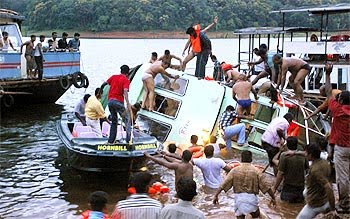
September 30, 2009; was a day of agony, sorrow and mourning for the people of Kerala,"The Gods Own Country." A tourist boat capsized in Thekkady lake, Kerala when 46 passengers drowned (nearly half of them were children) and lost their lives.The double decker boat ‘Jalakanyaka’ operated by Kerala Tourism Development Corporation (KTDC). capsized at a depth of 40 to 50 feet in Periyar Lake, at Periyar Wildlife Sanctuary, Thekkady around 17:30 hrs IST. It was one of the major accidents in the history of Kerala, and the authorities were not prepared to face such a tragedy as it was sudden and unexpected.
The accident happened when the boat "Jalakanyaka" tilted after several tourists moved to one side on sighting elephants on the banks of the lake and the driver lost control. This happened around sunset making rescue operations dfficult. It was shocking to note that none of the passen gers were wearing life jackets and there were no prior instructions to passengers on safety aspects by the boat crew before the journey. Also there were no life guards in the boat.
Drowning means death due to suffocation with or without aspiration of water while submerged in water.Near drowning means suffocation and asphyxia but with possible survival. Asphyxia due to submersion or hypoxia due to aspiration of water or a combination of both occur in drowning leading to death. Drowning can be either fresh water (well, lakes) or sea water.Also classified as dry or wet drowning. In dry drowning the victim is subjected to severe reflex laryngospasm and hypoxia following accidental contact with cold water and death is primarily due to asphyxia ( lungs are free of water). In wet drowning water enters the lung and cause alveolar flooding, alveolar edema, loss of surfactant, ventilation perfusion mismatch ,bronchospasm and hypoxia. Disruption of the alveolocapillary membranes lead on to ARDS.
The accident happened when the boat "Jalakanyaka" tilted after several tourists moved to one side on sighting elephants on the banks of the lake and the driver lost control. This happened around sunset making rescue operations dfficult. It was shocking to note that none of the passen gers were wearing life jackets and there were no prior instructions to passengers on safety aspects by the boat crew before the journey. Also there were no life guards in the boat.
Drowning means death due to suffocation with or without aspiration of water while submerged in water.Near drowning means suffocation and asphyxia but with possible survival. Asphyxia due to submersion or hypoxia due to aspiration of water or a combination of both occur in drowning leading to death. Drowning can be either fresh water (well, lakes) or sea water.Also classified as dry or wet drowning. In dry drowning the victim is subjected to severe reflex laryngospasm and hypoxia following accidental contact with cold water and death is primarily due to asphyxia ( lungs are free of water). In wet drowning water enters the lung and cause alveolar flooding, alveolar edema, loss of surfactant, ventilation perfusion mismatch ,bronchospasm and hypoxia. Disruption of the alveolocapillary membranes lead on to ARDS.
Wednesday, April 7, 2010
DILATED CARDIOMYOPATHY
A 7 year old boy is presented with chest discomfort and palpitation. He is also complaining of right iliac fossa pain.The pulse rate is 174/mt, BP 94/50 with mild elevated JVP. The monitor showed supraventricular tachycardia which responded to carotid sinus massage. He is febrile and has pallor.Chest is clear and CVS examination revealed an ejection systolic murmur of grade 2/4 radiated along left parasternal border and tachycardia. Abdominal examination showed Rt. iliac fossa tenderness with guarding.Lab findings were normal except for a high WBC count and Hb value of 9 Gms%. Previous records showed one ER admission with palpitation and the diagnosis was marked as suspected Cardiomyopathy(unclassified) with a probable viral etiology.The monitor again showed rapid pulse rate and the ECG is characteristic of supraventricular tachycardia with no response to carotid sinus massage this time.The patient was shifted to ICU for stabilisation, Oxygen by mask applied and cardiovascular status monitored. Inj. adenosine was given 2.6 mg iv and the heart rate slowed down initially to 100/mt but raising,so an immediate second bolus of adenosine 5.2 mg was given.Controlled fluid management started and temperature was maintained.The heart rate reduced to 132/mt and a second ECG showed SVT with QRS duration of .08 with QT 0.45 secs
The HGT was 5.5 and ABG showed mild respiratory alkalosis.Abdominal examination revealed acute appendicitis and was confirmed by ultrasonography.A decision to operate was made.The patient was administered amiodarone 5mg/kg and infusion 2.5 mic/kg/mt followed.The CXR showed cardiomegaly and an echocardiogram showed dilated left ventricle with global hypokinesia and impaired systolic function, posteriorly displaced mitral valve with MR, ejection fraction of 40%, all suggestive of a dilated cardiomyopathy.The heart rate is now stabilised on amiodarone and the cardiologist was consulted for the management of any intraoperative adverse events and to prepare for a temporary pacemaker insertion as an emergency intervention.Aspiration prophylaxis was given, Midazolam 1mg was given IV and the patient was shifted to operation theatre.
ECG, Oxygen saturation,Temperature ,NIBP etc were monitored. Anaesthesia was induced with IV ketamine 1 mg/kg and propofol 1mg/kg, along with IV fentanyl 1mcg/kg supplemented with oxygen, nitrous oxide and 1-1.5% isoflurane.Vecuronium was used to facilitate intubation at the dose of 1.5 mg/kg.(1) The heart rate and BP were stable throughout the procedure and a baseline infusion of amiodarone 1-2mic/kg/mt was on flow.Anaesthesia was maintained with O2/N2O in isoflurane (MAC 1-1.5) and intermittent boluses of vecuronium bromide IV, An arterial line and central venous catheter were inserted.TEE was not available. An esophageal stethescope was introduced. The capnogram waveforms are carefully observed.A central venous pressure of 6-8 cm of H2O was maintained by controlled fluid administration. Ideally a pulmonary artery catheter is indicated to monitor the filling pressures along with TEE. The reversal of neuromuscular block was achieved with neostigmine and glycopyrrolate and the postoperative period was uneventful.The child is stabilised on amiodarone 125 mg bid dose and was discharged with a maintenance dose of atenolol 25 mg so as to prevent the long term complications of amiodarone like thyroid dysfunction and pulmonary toxicity.
Dilated cardiomyopathy is characterised by left ventricular or biventricular dilatation and impaired ventricular contractility.The most common complication of dilated cardiomyopathy is progressive congestive cardiac failure.The commonest etiology is idiopathic or viral infections in children and alcohol abuse in adults.Most of the patients are asymptomatic with cardiomegaly and minimal CVS symptoms and present later in the course with cardiac failure, when the mortlity rate is high.The predictors of poor prognosis are(2) an ejection fraction of less than 0.25 (as seen on Echo, during the acute presentation of heart failure), left ventricular end diastolic dilatation, a hypokinetic left ventricle, the presence of mitral and tricuspid regurgitation
The goals (3) of anaesthetic management are
- Myocardial depression should be avoided
- Normovolemia is maintained
- To avoid overdose of drugs during induction as the circulation time is slow.
- Ventricular afterload is avoided
- Avoid sudden hypotension when regional anaesthesia is the choice
- Induction by midazolam/nitrous oxide/ vecuronium /isoflurane for GA
- Graded epidural anaesthesia with sedation using midazolam.The advantages are adequate post operative analgesia and less hemodynamic alteration.The changes in preload and afterload produced by epidural anesthesia mimic the pharmacological goals of treatment.(3) Here an anaesthetic level upto T4 is required and a well relaxed abdominal muscles are preferred.To achieve this goal the dosage of local anaesthetic requirement would be high which may precipitate sudden change in hemodynamics and the treatment of hypotension with ephedrine further worsens the CVS status.Phenylephrine if used to treat hypotension may cause increase in afterload which is detrimental.Hence the decision to administer GA.
1)Yamaguchi S, Wake K, Mishio M, et al. Anesthetic management of a patient with dilated cardiomyopathy under total intravenous venous anaesthesia with propofol and ketamine combined with continuous epidural analgesia. Masui 1999;48: 1232-34.
http://www.ncbi.nlm.nih.gov/pubmed/10586558
2 WILLIAM G, VALENTIN FUSTER: Idiopathic dilated Cardiomyopathy. New England Journal of Medicine; 331:1564-75, 1994.
3. ROBERT STOELTING K, STEPHEN F: Anesthesia And Co-Existing Disease, 4th Edition Lippincott-Raven; Ch. 7:117-120.
Wednesday, March 31, 2010
ANAPHYLAXIS THE EXTREME HYPERSENSITIVITY!
Anaphylaxis is an acute, severe, potentially life threatening allergic reaction (type 1 hypersensitivity) characterised by severe bronchospasm, angio neurotic edema and cardiovascular collapse following repeated exposure to an allergen to which the individual is already sensitised. Anaphylaxis can be caused by a variety of allergens.
Tuesday, March 23, 2010
SEPSIS ON THE GO.
Will you be careless about a lacerated sports injury or an unknown bite on your leg? Have you ever neglected a skin abrasion by a metal piece or thorn? Do insect bites need attention? Here is the story of a young labourer, a worker from a farm, who succumbed to death following sepsis due to an unattended injury/bite on his leg about which he was totally unaware.
This man was complaining of pain on his left hip and leg for two days.The left thigh appeared swollen with mild bluish discolouration on the anterolateral aspect.. He had no past history of any systemic illness and was in good health while joining the farm. Since 1 day he had low grade fever and there was progre ssive increase in difficulty of breathing. Examination showed tachypnea, dyspnea, Pulse: 122/mt, BP: 88/64 mm Hg, SPO2: 78% on room air, Pallor +, ABG: mild metabolic acidosis, Chest: b/l crepitations,CVS: tachycardia, CNS: alert and conscious, ABD: mild splenomegaly and abdominal wall edema, Swollen, warm and tender left leg with knee effusion and normal dorsalis pedis pulsation.A definite or identifiable mark of injury was not observed on the limb. A provisional diagnosis of cellulitis with septicemia or DVT with pulmonary embolism, was made. Patient was shifted to ICU for close observation of vitals and cardiorespiratory support. IV fluids started and oxygen by mask applied, dopamine 10-15 mic/kg/mt and dobutamine 5-10 mic/kg/mt were on flow. Blood investigations were ordered.Fluid aspirated from thigh and knee was serosanguinous in nature and awaited culture reports. Empirical broad spectrum antibiotics were started.Doppler ultrasound ruled out DVT or limb ischemia due to thrombus.X ray of left femur was normal.ECG showed sinus tachycardia, and CXR was normal. CT chest, abdomen and limb were considered but deferred as contrast could not be given due to derranged RFT, hypotension and abnormal coagulation profile. The echo cardiogram revealed Dilated left ventricle and impaired systolic function with EF 40%, and global hypokinesia, consistent with myocarditis.Fresh blood transfusion and FFP commenced. The blood investigations now showed
Hb 12.9 gm/dl, Platelet 100 K/UL, Derranged coagulation, RBS 120 mg/dl,CRP ++, ESR 70mm/hr, WBC 16K/uL, Uric acid 417 micmol/Lt.,LDH 820 u/l, CK 9495u/l, Liver function elevated bilirubin total and direct with high ALP and AST . RFT : creatinine 200 mic mol/ltr, Hypoalbuminemia, FDP 80 mic gram/ml, Urine Hemoglobin +++ and myoglobin + peripheral smear --> WBC more than 17% bands with toxic changes along with 10% atypical lymphocytes, Microcytic Hypochomic Anemia and Megakaryocytes
This man was complaining of pain on his left hip and leg for two days.The left thigh appeared swollen with mild bluish discolouration on the anterolateral aspect.. He had no past history of any systemic illness and was in good health while joining the farm. Since 1 day he had low grade fever and there was progre ssive increase in difficulty of breathing. Examination showed tachypnea, dyspnea, Pulse: 122/mt, BP: 88/64 mm Hg, SPO2: 78% on room air, Pallor +, ABG: mild metabolic acidosis, Chest: b/l crepitations,CVS: tachycardia, CNS: alert and conscious, ABD: mild splenomegaly and abdominal wall edema, Swollen, warm and tender left leg with knee effusion and normal dorsalis pedis pulsation.A definite or identifiable mark of injury was not observed on the limb. A provisional diagnosis of cellulitis with septicemia or DVT with pulmonary embolism, was made. Patient was shifted to ICU for close observation of vitals and cardiorespiratory support. IV fluids started and oxygen by mask applied, dopamine 10-15 mic/kg/mt and dobutamine 5-10 mic/kg/mt were on flow. Blood investigations were ordered.Fluid aspirated from thigh and knee was serosanguinous in nature and awaited culture reports. Empirical broad spectrum antibiotics were started.Doppler ultrasound ruled out DVT or limb ischemia due to thrombus.X ray of left femur was normal.ECG showed sinus tachycardia, and CXR was normal. CT chest, abdomen and limb were considered but deferred as contrast could not be given due to derranged RFT, hypotension and abnormal coagulation profile. The echo cardiogram revealed Dilated left ventricle and impaired systolic function with EF 40%, and global hypokinesia, consistent with myocarditis.Fresh blood transfusion and FFP commenced. The blood investigations now showed
Hb 12.9 gm/dl, Platelet 100 K/UL, Derranged coagulation, RBS 120 mg/dl,CRP ++, ESR 70mm/hr, WBC 16K/uL, Uric acid 417 micmol/Lt.,LDH 820 u/l, CK 9495u/l, Liver function elevated bilirubin total and direct with high ALP and AST . RFT : creatinine 200 mic mol/ltr, Hypoalbuminemia, FDP 80 mic gram/ml, Urine Hemoglobin +++ and myoglobin + peripheral smear --> WBC more than 17% bands with toxic changes along with 10% atypical lymphocytes, Microcytic Hypochomic Anemia and Megakaryocytes
Saturday, March 13, 2010
GUILLAIN-BARRE SYNDROME
Clinical Manifestations
- Rapidly evolving, progressive, ascending motor paralysis with or without sensory disturbance and with areflexia
- Lower cranial nerves are frequently involved and presented with difficulty in swallowing and maintaining airway
- Bulbar involvement is most frequently manifested as B/L facial paralysis
- Fever and other constitutional symptoms are usually absent
- Diminished DTR with absent proprioception. There may be marked sensory loss
- Bladder dysfunction is late and transient.
- Deep aching pain in muscles and back
- Difficulty in swallowing due to pharyngeal muscle weakness and impaired ventilation due to intercostal muscle paralysis in 30% of cases and they require ventilatory support
- Marked autonomic dysfunction leads to Wide fluctuations in blood pressure – hypo / hyper tension, Sudden profuse diaphoresis,Peripheral vasoconstriction Resting tachycardia,Cardiac conduction abnormality, Orthostatic hypotension – severe, Thromboembolism due to immobilisation, & sudden death
- Metabolic derangements -Hyponatremia due to excessive ADH secretion
Friday, March 12, 2010
PREGNANT PATIENT WITH DVT IN PAIN
24 year old second gravida with 9 weeks of pregnancy reports to the emergency department with 1 day history of pain left loin spreading to left lower limb.The patient gives history of contraceptive injections. On examination the left lower limb appeared swollen and tender. The homan's sign was positive.Femoral and Popleateal pulsations were felt but dorsalis pedis feebly felt compared to right leg. The limb was not pale or cold. A provisional diagnosis of DVT is made and a venous doppler was ordered which showed DVTof the left lower limb extending upto left ileofemoral vein.
Patient is admitted in icu, heparin infusion started 5000units iv and then 1000units/hr keeping the APTT level 1.5 to 2 times than normal.Monitoring of Spo2, ECG, NIBP and temperature commenced. Serial ABG s were performed to asses the respiratory status. Lower limb mesurements were taken frequently and observed for development of compartmental syndrome.Arterial doppler showed no arterial insufficiency and echocardiogram showed normal study. The CXR also was normal
Anesthetic referral was sent for pain relief.
Providing pain relief to this patient was a real challenge before the anaesthetist He has to consider the following things before formulating the therapy
1) Patient is on thrombolytic therapy
2)Teratogenecity of anaesthetic agents
3)Effect of analgesic agents on uterine tone
4)Need of careful monitoring of the respiratory system as chance of pulmonary embolism is high.
NSAIDS including diclofenac sodium
Paracetamol
Paracetamol is considered to be safe and can be given intramuscular injection in case of
severe pain. But the efficacy is limited as continuous pain relief is not possible.The effects on maternal Liver like fatty liver or liver failure are of concern but are rare and occurs only on long term treatment.IV preparation is preferred as deep IM can cause hematoma formation as patient is on heparin
Patient is admitted in icu, heparin infusion started 5000units iv and then 1000units/hr keeping the APTT level 1.5 to 2 times than normal.Monitoring of Spo2, ECG, NIBP and temperature commenced. Serial ABG s were performed to asses the respiratory status. Lower limb mesurements were taken frequently and observed for development of compartmental syndrome.Arterial doppler showed no arterial insufficiency and echocardiogram showed normal study. The CXR also was normal
Anesthetic referral was sent for pain relief.
Providing pain relief to this patient was a real challenge before the anaesthetist He has to consider the following things before formulating the therapy
1) Patient is on thrombolytic therapy
2)Teratogenecity of anaesthetic agents
3)Effect of analgesic agents on uterine tone
4)Need of careful monitoring of the respiratory system as chance of pulmonary embolism is high.
NSAIDS including diclofenac sodium
Paracetamol
Paracetamol is considered to be safe and can be given intramuscular injection in case of
severe pain. But the efficacy is limited as continuous pain relief is not possible.The effects on maternal Liver like fatty liver or liver failure are of concern but are rare and occurs only on long term treatment.IV preparation is preferred as deep IM can cause hematoma formation as patient is on heparin
Thursday, March 11, 2010
ANAESTHESIA IN DENTAL CHAIR
As a rule patients should be premedicated with atropine to reduce oral secretions and to reduce the occurrence of vasovagal syncope.(In children this may be administered at the dose of 0.02mg/kg IV following induction and iv line placement) In a co operative child the IV cannula may be secured after topical application of EMLA cream also. Intravenous induction with Propofol and lidocaine may be condsidered for children above 3 yrs. A nasal mask or LMA may be used to protect the airway with additional pre op oral packing, particularly when nasal mask is used for delivering inhalational anaesthesia. Maintenance of anaesthesia is continued with oxygen, nitrous oxide with isoflurane allowing spontaneous ventilation. Alternatively inhalation induction with sevoflurane or halothane is preferred for children less than 3 yrs.The eyes should be protected. For maintenance, propofol or sevoflurane may be continued. Additional analgesia may be obtained by local nerve blocks by surgeon. If possible the child my be placed in the lateral position keeping the head low with proper stabilisation of head and neck.Use of nitrous oxide may be reduced or stopped with concurrent administration of short acting analgesics like fentanyl, to prevent hypoxia,provide analgesia and to make available 100% oxygen. Post op analgesia can be achieved with paracetamol or diclofenac suppositories or with intramuscular opioids.
Another technique called inhalational sedation is used for adults who are unwilling for local anaesthesia. Here conscious sedation is given using iv anaesthetic agents followed by analgesia supplemented with local anaesthetics once the sedative effect is established.Patients are given slow incremental doses of midazolam with oxygen and less than 50% nitrous oxide.A soft but weighted mask is put over the nostrils. It is essential that patient is conscious throughout the procedure and can control his airway. The patient should be able to communicate with the surgeon during the procedure. Mouth props are discouraged as they mask the patients inability to keep the mouth open, - an ominous sign of oversedation. Standard monitoring include NIBP, ECG, Temperature probe and Pulse Oximetry. Patient should be given 100 % oxygen following the procedure to avoid diffusion hypoxia and flumazenil should be available to reverse the residual effect of benzodiazepines effect, if required.
When to think of intubation?
- Difficult access as in impacted tooth, macroglossia, short neck
- Excessive uncontrolled bleeding
- maxillofacial or major dental surgery
- Mentally handicapped
- Obstruction of nasal passages, large adenoids where nasal mask is not effective
Problems in dental chair
- Reduced venous return, so more severe reaction to a vasovagal attack due to pain
- Increased chance of air embolism in sitting position
- Unprotected airway ie shared airway between surgeon and anaesthetist
- Aspiration of blood or mucus can cause laryngospasm,
- Adrenaline in local anaesthetic can cause arrhythmias in presence of halothane
- Higher incidence of arrhythmias due to stimulation of 5 th cranial nerve
- Nasal bleeding if nasal airway is used to deliver the anaesthetic
- Fainting due to cerebral hypoxia or cardiac arrest may be unrecognised
- Difficulty in initiating CPCR once cardiac arrest occurs
- Foreign body obstruction of the airway by needles or dentures,necessitating removal by bronchoscopy
- Uncontrolled and profuse bleeding into the airway in patients not screened for coagulopathies especially in out patients
- Higher incidence of infective endocarditis
- Anaphylaxis, to local anaesthetics or other drugs
- Malignant hyperpyrexia if halothane is used
- Difficulty in administering post op analgesia
Monday, March 8, 2010
MUSCLE RELAXANTS IN THE ICU.
Muscle relaxants are used in icu to facilitate mechanical ventilation of the critically ill patients.The drugs commonly used are pancuronium, vecuronium, atra curium, cisatra curium, and rocuronium. Muscle relaxants are essential in the management of tetanus, status epilepticus, to provide hyper ventilation in head injury, or after precise surgical intervention like tracheal anastomosis and vascular anastomosis.These agents are also used to reduce oxygen consumption or to reduce work of breathing in COPD and ARDS(where inflation pressures are high) patients.
Apart from the beneficial effects the complications of these agents are also of concern and are investigated. These can be classified as complications due to short term use and long term use
Short term use
- Side effects atracurium causing histamine release or hyperkalemia due to suxamethonium
- Inadequate analgesia or sedation as these components are under estimated when relaxants are used
- Immobility : DVT decubitus ulcers, peripheral nerve injuries.
- Inability to cough: retained secretions, atelectasis and infection
- Persistent paralysis on stoppage of the drugs "Critical Illness Neuropathy and Critical Illness Myopathy", Steroid induced myopathy.
- CNS effects of drugs eg: laudanosine causing convulsions
Wednesday, March 3, 2010
H1N1 AND HAND HYGIENE
There is a raising concern and anxiety about the spread of H1N1 influenza among people and health care personnel, as the number of reported cases are on the hike. Eventhough the mode of transmission is by droplet spread, transmission through conta minated hands are also considered to be significant. Along with personal protection devices like N-95 mask, goggles, apron, and gloves the importance of hand hygiene is also stressed, as prevention is the major goal for the control of spread. Several studies conducted on this issue have proved that practise of safe and appropriate hand hygiene is an important public health initiative to reduce pandemic H1N1 influenza transmission.
The commercially available hand sanitisers are alcohol based with the addition of various antiseptics and emollients.They are available both in liquid form and gel form. The advantages of alcohol based sanitisers are.
Research by (Ref), Grayson ML, Melvani S, Druce J, Barr IG, Ballard SA, Johnson PD, Mastorakos T, Birch C., Infectious Diseases Department, Austin Health, Heidelberg, Victoria 3084, Australia, showed that hand hygiene with soap and water or alcohol-based hand rub is highly effective in reducing influenza A virus on human hands, although washing with soap and water is the most effective intervention.
Following the" NO FEAR, FIGHT INFLUENZA" campaign, instant hand sanitisers are made available everywhere, including supermarkets,restaurants, clinics, schools, factories etc.One danger of alcohol based sanitisers is that they are flammable and can cause burns.It happened in one hospital where the hospital cleaner used hand wash gel after cleaning the toilets and immediately lighted up a cigarette, holding up his hands to protect the flame from the wind, and his hands burst into flames.Anoher incidence reported from a birthday party where the children were playing with a sanitiser spray and their dress caught fire from candles. So it is advised that after sanitiser use one should make sure that the hands are dry before lighting gas stove,cigarettes or candles and when handling fire or dealing with any job which produces spark for example welding works.
Agents that reduce skin microflora are called antiseptics, whereas agents that reduce microflora on non living objects are called disinfectants. Antibiotic creams or dettol(chlorhexidine) are considered antiseptics, while glutaraldehyde (cidex) or floor cleaners like lysol(phenol based) are categorised as disinfectants. The efficacy of disinfectants or antiseptics are determined by Rideal Walker coefficient and is obtained by dividing the figure indicating the degree of dilution of the disinfectant that kills a microorganism in a given time by that indicating the degree of dilution of phenol that kills the organism in the same space of time under similar conditions.
Ref: Clin Infect Dis. 2009 Feb 1;48(3):285-91.
Watch a video on H1N1
The commercially available hand sanitisers are alcohol based with the addition of various antiseptics and emollients.They are available both in liquid form and gel form. The advantages of alcohol based sanitisers are.
- Instant in action , effective in less than 15 seconds
- Reliable, almost 99% of pathogens including viruses are killed(actiton on spores doubtful)
- Easy to use as it immediately dries up and hand washing is not necessary
- leaves less stain than other agents
Research by (Ref), Grayson ML, Melvani S, Druce J, Barr IG, Ballard SA, Johnson PD, Mastorakos T, Birch C., Infectious Diseases Department, Austin Health, Heidelberg, Victoria 3084, Australia, showed that hand hygiene with soap and water or alcohol-based hand rub is highly effective in reducing influenza A virus on human hands, although washing with soap and water is the most effective intervention.
Following the" NO FEAR, FIGHT INFLUENZA" campaign, instant hand sanitisers are made available everywhere, including supermarkets,restaurants, clinics, schools, factories etc.One danger of alcohol based sanitisers is that they are flammable and can cause burns.It happened in one hospital where the hospital cleaner used hand wash gel after cleaning the toilets and immediately lighted up a cigarette, holding up his hands to protect the flame from the wind, and his hands burst into flames.Anoher incidence reported from a birthday party where the children were playing with a sanitiser spray and their dress caught fire from candles. So it is advised that after sanitiser use one should make sure that the hands are dry before lighting gas stove,cigarettes or candles and when handling fire or dealing with any job which produces spark for example welding works.
Agents that reduce skin microflora are called antiseptics, whereas agents that reduce microflora on non living objects are called disinfectants. Antibiotic creams or dettol(chlorhexidine) are considered antiseptics, while glutaraldehyde (cidex) or floor cleaners like lysol(phenol based) are categorised as disinfectants. The efficacy of disinfectants or antiseptics are determined by Rideal Walker coefficient and is obtained by dividing the figure indicating the degree of dilution of the disinfectant that kills a microorganism in a given time by that indicating the degree of dilution of phenol that kills the organism in the same space of time under similar conditions.
Ref: Clin Infect Dis. 2009 Feb 1;48(3):285-91.
Watch a video on H1N1
Monday, March 1, 2010
AIRWAY MANAGEMENT IN A PATIENT WITH HUGE THYROID
Airway management of a patient with a huge thyroid swelling is a real challenge to the anaesthetist. The anaesthetic concerns and the management of airway are discussed here.
60 year old female patient is admitted to the A&E department with history of neck swelling and stridor. She is a known case of medullary carcinoma thyroid with lymph node metastases.
The patient assumed a sitting position as stridor exacerbated on lying supine and looks distressed .She was oriented conscious and responding to comm ands. Examination of the chest revealed b/l basal creps and oxygen saturation was 88% with oxygen mask. ABG showed resp acidosis with hypoxemia.The pulse rate was regular and ECG was normal.The thyroid swelling was huge and completely occupying the anterior neck with the trachea not at all palpable at any point and with evidence of possible retrosternal extension.Oxygen by mask administered and patient postioned sitting, leaned forward.
Investigations done:
Additional investigations (may be deferred here as this patient requires emergency airway access)
1) Induction or any anaesthetic intervention should be done with the patient in sitting position, as supine position further compromises airway.
2)Emergency tracheostomy, minintracheostomy or retrograde tracheostomy cannot be
performed here due to tumour extension.
3)Distorted airway anatomy makes intubation impossible even with fiber optic bronchoscopy.
4)Ventilation through Fastrach may not be adequate and may not be effective due to tracheal compression, if intubation through Fastrach fails.
5)Airway blocks like trans tracheal or superior laryngeal nerve blocks cannot be performed.
6) Tracheostomy malfunction may occur post operatively due to tracheal collapse because of tracheomalacia.
The ideal management in this case would be a fiberoptic trial of intubation through the nasal or oral route, in the sitting position with topical airway anaesthesia, followed by palliative debulking of the tumor and then tracheostomy.
following rigid fiber optic bronchoscopy and inhalation induction using sevoflurane in the sitting postition followed either by blind intubation or fiberoptic aided intubation.
Anaesthetist should be vigilant, careful and well prepared while dealing with neck masses compromising airway. Management should done in operation theatre set up with all available equipments ready to tackle the difficult situation. The ENT and General surgeon also should be available to deal with emergency.
Thanx to Dr.Santhosh Kumar Sathiarajan, Radiologist, Ibri hospital, Muscat.
Comments? Click the comment tab below
60 year old female patient is admitted to the A&E department with history of neck swelling and stridor. She is a known case of medullary carcinoma thyroid with lymph node metastases.
The patient assumed a sitting position as stridor exacerbated on lying supine and looks distressed .She was oriented conscious and responding to comm ands. Examination of the chest revealed b/l basal creps and oxygen saturation was 88% with oxygen mask. ABG showed resp acidosis with hypoxemia.The pulse rate was regular and ECG was normal.The thyroid swelling was huge and completely occupying the anterior neck with the trachea not at all palpable at any point and with evidence of possible retrosternal extension.Oxygen by mask administered and patient postioned sitting, leaned forward.
Investigations done:
- Routine hemogram
- CXR showed severe narrowing and deviation of the trachea to the right side, by compression from the mass
- CT scan showed narrowed tracheal orifice(the black round hole) with compression of the esophagus and nodular metastases
Additional investigations (may be deferred here as this patient requires emergency airway access)
- Pulmonary function tests may show a restrictive defect with high FEV1/FVC ratio
- Carotid angiogram to assess the vessels
1) Induction or any anaesthetic intervention should be done with the patient in sitting position, as supine position further compromises airway.
2)Emergency tracheostomy, minintracheostomy or retrograde tracheostomy cannot be
performed here due to tumour extension.
3)Distorted airway anatomy makes intubation impossible even with fiber optic bronchoscopy.
4)Ventilation through Fastrach may not be adequate and may not be effective due to tracheal compression, if intubation through Fastrach fails.
5)Airway blocks like trans tracheal or superior laryngeal nerve blocks cannot be performed.
6) Tracheostomy malfunction may occur post operatively due to tracheal collapse because of tracheomalacia.
The ideal management in this case would be a fiberoptic trial of intubation through the nasal or oral route, in the sitting position with topical airway anaesthesia, followed by palliative debulking of the tumor and then tracheostomy.
- Lignocaine jelly or gargle may be used to anaesthetise the posterior tongue
- Glossopharyngeal nerve block can be performed intra orally by injecting local anaesthetic into the base of the palatoglossal fold.
- Nasal passages may be anaesthetised by lignocaine jelly using swabs or catheter.
- Excellent anaesthesia of the larynx and trachea can be achieved with topical aerosolised lignocaine.
- Intravenous sedation can be given with midazolam or fentanyl in titrated doses during the procedure, if patient is uncomfortable.
- A smaler sized re inforced tube is preferred for intubation
following rigid fiber optic bronchoscopy and inhalation induction using sevoflurane in the sitting postition followed either by blind intubation or fiberoptic aided intubation.
Anaesthetist should be vigilant, careful and well prepared while dealing with neck masses compromising airway. Management should done in operation theatre set up with all available equipments ready to tackle the difficult situation. The ENT and General surgeon also should be available to deal with emergency.
Thanx to Dr.Santhosh Kumar Sathiarajan, Radiologist, Ibri hospital, Muscat.
Comments? Click the comment tab below
Thursday, February 25, 2010
NITRIC OXIDE: THE SAVIOUR.
Nitric oxide (NO) is one of the nitrogen oxides and is synthesized within cells by an enzyme NO synthase. It was formerly known as endothelium-derived relaxing factor (EDRF). Being one of the fundamental mediators in physiological processes, this enzyme catalyses the oxidation of L-arginine to L-citrulline, producing NO, which diffuses into vascular smooth muscle, activating guanylate cyclase which in turn converts guanosine triphosphate into cyclic guanosine monophosphate (cGMP),causing vascular relaxation.
Nitric oxide synthetase is present in two forms a)The constitutive form (eNOS), which is present in vascular, neuronal, cardiac tissue, skeletal muscle and platelets, producing small quantities of NO continuously. Here NOS is Ca2+/calmodulin dependant and is stimulated by cGMP.and b)the inducible form (iNOS),present in endo thelium, myocytes, macrophages and neutrophils, which produces relatively large quatities of NO after exposure to endotoxins in sepsis.
Biological effects of Nitric oxide ;
• Vascular endothelium: producing vascular relaxation.
• Platelets: involved in aggregation and adhesion of platelets
• Brain tissue: acts as a neurotransmitter.
• Macrophages: involved in the response to infection.
Nitric oxide synthetase is present in two forms a)The constitutive form (eNOS), which is present in vascular, neuronal, cardiac tissue, skeletal muscle and platelets, producing small quantities of NO continuously. Here NOS is Ca2+/calmodulin dependant and is stimulated by cGMP.and b)the inducible form (iNOS),present in endo thelium, myocytes, macrophages and neutrophils, which produces relatively large quatities of NO after exposure to endotoxins in sepsis.
Biological effects of Nitric oxide ;
• Vascular endothelium: producing vascular relaxation.
• Platelets: involved in aggregation and adhesion of platelets
• Brain tissue: acts as a neurotransmitter.
• Macrophages: involved in the response to infection.
Monday, February 22, 2010
RESUSCITATION IN PREGNANCY DURING AND AFTER
The overall incidence of cardiac arrest during late pregnancy is estimated to be one in 30000, however survival rate following such an event is extremely low. Most deaths are due to acute medical conditions associated with pregnancy rather than due to causes from pregnancy itself. Resusctation in pregnancy is different as we have two potential patients, the mother and the fetus. The best hope of fetal survival is maternal survival. Factors peculiar to pregnancy such as the anatomical, physiological and pathological changes make resuscitation difficult in late pregnancy. The major considerations are :
Respiratory
2) The abc of resuscitation, BMJ
Respiratory
- Increased alveolar ventilaton and oxygen consumption
- Increased oxygen demand
- Reduced chest compliance
- Reduced functional residual capacity
- Increased risk of aspiration
- Laryngeal edema
- Airway obstruction due to edema and soft tissue hypertrophy
- Incompetent gastroesophageal (cardiac) sphincter
- Increased intragastric pressure
- Increased risk of regurgitation
- Increased acidity of gastric contents
- Physiological anemia
- Supine hypotension syndrome
- Obesity, turning the patient to one side is difficult and requires assistance
- Hypertrophied breasts cause difficulty in laryngoscopy and prevents effective chest compression or defibrillation
- Remove dentures or foreign body from airway
- Suction and artificial airway, if available
- Ventilation by mouth to mouth or using an ambu bag. Remember to apply cricoid pressure
- Turn the patient to get lateral displacement of uterus and thereby preventing venacaval obstruction by the gravid uterus. A left lateral displacement is made by tilting the body to 30 degrees which can be achieved with a cardiff wedge kept beneath the right buttock or a rolled towel or pillow if wedge is not available. The other techniques to provide tilt are by manual displacement of uerus or by tilting onto the back of an upturned chair. A human wedge is the one where the patient is turned onto a rescuers knees to provide a stable position for BLS.
- Chest compressions at the rate of 30:2, consider the cephalad movement of the diaphragm, and so the hands should be kept at a higher level during compression
- Early inyubation is recommended
- Airway is "difficult"
- Tracheal intubation difficult due to anatomical changes like short neck, edematous soft tissues, large tongue, and large breasts
- So isertion of laryngoscope is difficult hence, short handle may be used for the scope, removing the blade and re attach after insertion into the oral cavity, or using a curved blade like Mc Coy, or polio blade etc are advised
- LMA classic or FAST TRACH are advised for emergency airway access if intubation fails. Release cricoid pressure during insertion and re apply after insertion of LMA eventhough cricoid pressure is not very effective with LMA insitu.
- Defibrillation when needed should be done according to standard ACLS defibrillation doses. Remove all fetal or maternal monitors while giving shock. DC shocks are found to be not harmful to the fetus
- Immediate iv access and fluid resuscitation in case of hemorrage
- Consider low tidal volumes, enough to make a visible chest rise
- Consider early caesarean section when standard rsuscitative measures fail to restore maternal circulation and shoul be done in less than 5 minutes.(even after mother's heart stops) caesarean section helps to relieve aortocaval compression and incrase venous return, thereby improve cardiac output.It also increases thoracic compliance
- Vaso pressors are also used as per standard ACLS protocols eventhough fetal blood flow is reduced
- Immediate abdominal ultrasound for concealed hemorrage
- Excess magnesium sulphate(may be iatrogenic). Administer calcium gluconate 1 gram or one ampoule. Empirical therapy also is life saving.
- Acute coronary syndromes to be considered in elder patients. Fibrinolytics are contra indicated hence percutaneous coronary intervention is the treatment of choice for STEMI
- Pre eclampsia, or eclampsia, consider treatment of hypertension or emergency caesarean section
- Aortic dissection, a rare possibility
- Life threatening pulmonary embolism or stroke here fibrinolytics must be administered as prognosis was found to be good ,in spite of risk, in massive embolus
- Amniotic fluid embolism consider immediate CABG
- Trauma and drug overdose
2) The abc of resuscitation, BMJ
Subscribe to:
Posts (Atom)