DISCLAIMER

The contents of this blog are solely meant for information & education purpose only.These may be the basis of actual treatment, but not necessarily. Information from other websites and journals are also included. So the author is not responsible for any inaccuracy,loss, or damage that may arise due to the use of these informations published here. I do respect copyright & always give credits to the original author(s) and thankful to them. Inspite of my utmost effort and care there can be human error. If anyone finds any violation of copyright please inform me at anesthesiatoday@gmail.com and necessary action will be taken soon as possible.
My blog is also non-commercial.


Wednesday, April 28, 2010

DIFFUSION HYPOXIA

More than pain, he was worried of the disfigurement it caused.A small nevus just above the right eyebrow was a source of worry and disappointment which made him think of a cosmetic repair for better appearance. Mathew Davids,a 38 year old software engineer from India and employed in USA flew to his home country eventhough one of the US hospitals (with the best facil ities in world) offered a cosmetic cum plastic repair for him within affordable cost.He thought he will be more comfortable amidst of friends and relatives.

After 2 days he was scheduled for surgery. The pre anaesthetic check went smooth except for an elevated diastolic BP record of 94 mmHg. He was given GA spontaneous following propofol induction and fentanyl infusion using classic LMA.  Maintenance of anaesthesia was with 4-6% sevoflurane in oxygen and nitrous oxide.Intra operative BP, SPO2, ETCO2 and HR were within normal limits. Surgery completed in 32 minutes, LMA removed and he was shifted to recovery room.He was drowsy but arousable.

The recovery nurse who was busy with the management of some major post operative cases thought that his surgery is too short and doesn't require intensive monitoring.The oxygen mask was applied to his face but the other end of the tubing got disconnected on patient movement and was un noticed

There is a fall in saturation and SPO2 low alarm on monitor alerted the staff nurse. She rushed to his trolley and found him unresponsive and cyanosed.The monitor showed severe bradycardia and ST elevation on ECG. Immediate mask ventilation, CPCR  and intubation followed.Patient was shifted to ICU where a 12 lead ECG showed STEMI and the troponin value was raised.A decision to thrombolyse was made and initiated. He was on ventilator for three days and got discharged without any neurological deficit.The probable cause for this incidence was thought to be diffusion hypoxia with hypercarbia.Who is the cul prit here? The anaesthesiologist who kept the patient in deep inhalational plane maintaining spontaneous ventilation for 32 minutes or the recovery nurse who neglected the importance of administering oxygen for a GA case?

Diffusion hypoxia (fink effect) means outpouring of large volumes of nitrous oxide into the lung during recovery from general anaesthesia and subsequent hypoxia.This is due to
1) Direct replacement of oxygen from lung
2) Diluting alveolar carbon dioxide causing decreased drive for ventilation.
    This effect is seen in first 5-10 minutes of recovery when large volumes of nitrous oxide is released into the lung.The blood gas solubility of nitrous oxide is 0.42 which is less than that of any other inhalational agents.So rapid alveolar concentration following inhalation.But N2O is more soluble than nitrogen in blood. Hence blood and body fluids are rich in N2O.At the end of anaesthesia N2O diffuses back into alveoli from blood down a concentration gradient and this diffusion back is rapid than uptake of nitrogen from the alveoli by blood. This leads to replacement of all alveolar gases by nitrous oxide and subsequent hypoxia.

    Fig. showing relationship between O2 and N2O following recovery.

    So it is mandatory that 100% oxygen should be given for all GA cases following recovery for about 5-10 minutes to avoid the occurrence of diffusion hypoxia

    Ref: 1) Millers Anaesthesiology 7th edition
            2John L.Bezzant,M.D, http://library.med.utah.edu/kw/derm/nitrous/05ni.htm

    Monday, April 19, 2010

    CELIAC PLEXUS BLOCK

    Celiac plexus lies anterior to the aorta at the level of the first lumbar vertebra, between the origin of celiac and superior mesenteric arteries The adre nal glands lie lateral to the plexus and  stomach and pancreas lie anteriorly. The connections include Preganglionic sympathetic fibres from splanchnic nerves, Preganglionic parasympathetic from vagus, Sensory fibres from phrenic and vagus, Afferent fibres concerned with nociception
    The three pairs of splanchnic nerves descending to the celiac plexus are
     1) Greater splanchnic nerves from T5-T9
     2) Lesser splanchnic nerves from T10 and T11 segments
     3) Least splanchnic from T12

    The three pairs of ganglia in the plexus are 
     1)Celiac ganglia
      2)Superior mesenteric ganglia
      3)Aorticorenal ganglia
    The nociceptive afferent fibres travel from viscera along with the sympathetic fibres, through the ganglia, splanchnic nerve, sympathetic chain, white rami communicans and then synapse in the dorsal root ganglia.The proximal axon of these bodies synapse in the dorsal horn of the spinal cord

    The blockade of the celiac pleexus causes blockade of pain transmission from the visceral structures including Pancreas, Liver, Gall bladder, Omentum, Mesentery, Alimentary tract, and complete blockade of the Sympathetic fibres causing increased Parasympathetic activity manifested as increased intestinal motility and relaxed sphincters. The sympathetic blockade to the splanchnic vessels cause vasodialatation and hypotension

    Agents used for blockade are 0.5% bupivacaine with adrenaline1:200000 around 30ml, 15 ml on either side with or without steroids(dexamethasone) for chronic pain. Neurolytic blockade is indicated in abdominal malignancies where alcohol 50-100% or 10% phenol is used. The pain on injection of alcohol can be minimised with combination of bupivacaine 1:1 ratio

    Wednesday, April 14, 2010

    THE 'JALAKANYAKA TRAGEDY'

    September 30, 2009; was a day of agony, sorrow and mourning for the people of Kerala,"The Gods Own  Country." A tourist boat capsized in Thekkady lake, Kerala when 46 passengers drowned  (nearly half of them were children) and lost their lives.The double decker boat ‘Jalakanyaka’ operated by Kerala Tourism Development Corporation (KTDC). capsized at a depth of 40 to 50 feet in Periyar Lake, at Periyar Wildlife Sanctuary, Thekkady around 17:30 hrs IST. It was one of the major accidents in the history of Kerala, and the authorities were not prepared to face such a tragedy as it was sudden and unexpected.

    The accident happened when the boat "Jalakanyaka" tilted after several tourists moved to one side on sighting elephants on the banks of the lake and the driver lost control. This happened around sunset making rescue operations dfficult. It was shocking to note that none of the passen gers were wearing life jackets and there were no prior instructions to passengers on safety aspects  by the boat crew before the journey. Also there were no life guards in the boat.


    Drowning means death due to suffocation with or without aspiration of water while submerged in water.Near drowning means suffocation and asphyxia but with possible survival. Asphyxia due to submersion or hypoxia due to aspiration of water or a combination of both occur in drowning leading to death. Drowning can be either fresh water (well, lakes) or sea water.Also classified as dry or wet drowning. In dry drowning the victim is subjected to severe reflex laryngospasm and hypoxia following accidental contact with cold water and death is primarily due to asphyxia ( lungs are free of water). In wet drowning water enters the lung and cause alveolar flooding, alveolar edema, loss of surfactant, ventilation perfusion mismatch ,bronchospasm and hypoxia. Disruption of the alveolocapillary membranes lead on to ARDS.

    Wednesday, April 7, 2010

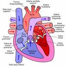
    DILATED CARDIOMYOPATHY

    A 7 year old boy is presented with chest discomfort and palpitation. He is also complaining of right iliac fossa pain.The pulse rate is 174/mt, BP 94/50 with mild elevated JVP. The monitor showed supraventricular tachycardia which responded to carotid sinus massage. He is febrile and has pallor.Chest is clear and CVS examination revealed an ejection systolic murmur of grade 2/4 radiated along left parasternal border and tachycardia. Abdominal examination showed Rt. iliac fossa tenderness with guarding.Lab findings were normal except for a high WBC count and Hb value of 9 Gms%. Previous records showed one ER admission with palpitation and the diagnosis was marked as suspected Cardiomyopathy(unclassified) with a probable viral etiology.The monitor again showed rapid pulse rate and the ECG is characteristic of supraventricular tachycardia with no response to carotid sinus massage this time.The patient was shifted to ICU for stabilisation, Oxygen by mask applied and cardiovascular status monitored. Inj. adenosine was given 2.6 mg iv and the heart rate slowed down initially to 100/mt but raising,so an immediate second bolus of adenosine 5.2 mg was given.Controlled fluid management started and temperature was maintained.The heart rate reduced to 132/mt and a second ECG showed SVT with QRS duration of .08 with QT 0.45 secs

    The HGT was 5.5 and ABG showed mild respiratory alkalosis.Abdominal examination revealed acute appendicitis and was confirmed by ultrasonography.A decision to operate was made.The patient was administered amiodarone 5mg/kg and infusion 2.5 mic/kg/mt followed.The CXR showed cardiomegaly and an echocardiogram showed dilated left ventricle with global hypokinesia and impaired systolic function, posteriorly displaced mitral valve with MR, ejection fraction of 40%, all suggestive of a dilated cardiomyopathy.The heart rate is now stabilised on amiodarone and the cardiologist was consulted for the management of any intraoperative adverse events and to prepare for a temporary pacemaker insertion as an emergency intervention.Aspiration prophylaxis was given, Midazolam 1mg was given IV and the patient was shifted to operation theatre.

    ECG, Oxygen saturation,Temperature ,NIBP etc were monitored. Anaesthesia was induced with IV ketamine 1 mg/kg and propofol 1mg/kg, along with IV fentanyl 1mcg/kg supplemented with oxygen, nitrous oxide and 1-1.5% isoflurane.Vecuronium was used to facilitate intubation at the dose of 1.5 mg/kg.(1) The heart rate and BP were stable throughout the procedure and a baseline infusion of amiodarone 1-2mic/kg/mt was on flow.Anaesthesia was maintained with O2/N2O in isoflurane (MAC 1-1.5) and intermittent boluses of vecuronium bromide IV, An arterial line and central venous catheter were inserted.TEE was not available. An esophageal stethescope was introduced. The capnogram waveforms are carefully observed.A central venous pressure of 6-8 cm of H2O was maintained by controlled fluid administration. Ideally a pulmonary artery catheter is indicated to monitor the filling pressures along with TEE. The reversal of neuromuscular block was achieved with neostigmine and glycopyrrolate and the postoperative period was uneventful.The child is stabilised on amiodarone 125 mg bid dose and was discharged with a maintenance dose of atenolol 25 mg so as to prevent the long term complications of amiodarone like thyroid dysfunction and pulmonary toxicity.

    Dilated cardiomyopathy is characterised by left ventricular or biventricular dilatation  and  impaired ventricular contractility.The most common complication of dilated cardiomyopathy is progressive congestive cardiac failure.The commonest etiology is idiopathic or viral infections in children and alcohol abuse in adults.Most of the patients are asymptomatic with cardiomegaly and minimal CVS symptoms and present later in the course with cardiac failure, when the mortlity rate is high.The predictors of poor prognosis are(2) an ejection fraction of less than 0.25 (as seen on Echo, during the acute presentation of heart failure), left ventricular end diastolic dilatation, a hypokinetic left ventricle, the presence of mitral and tricuspid regurgitation

    The goals (3) of anaesthetic management are
    • Myocardial depression should be avoided
    • Normovolemia is maintained
    • To avoid overdose of drugs during induction as the circulation time is slow.
    • Ventricular afterload is avoided
    • Avoid sudden hypotension when regional anaesthesia is the choice
    Alternate anaesthetic techniques are
    • Induction by midazolam/nitrous oxide/ vecuronium  /isoflurane for GA
    • Graded epidural anaesthesia with sedation using midazolam.The advantages are adequate post operative analgesia and less hemodynamic alteration.The changes in preload and afterload produced by epidural anesthesia mimic the pharmacological goals of treatment.(3) Here an anaesthetic level upto T4 is required and  a well relaxed abdominal muscles are preferred.To achieve this goal the dosage of local anaesthetic requirement would be  high which may precipitate sudden change in hemodynamics  and the treatment of hypotension with ephedrine further worsens the CVS status.Phenylephrine if used to treat hypotension may cause increase in afterload which is detrimental.Hence the decision to administer GA.
    Ref:
    1)Yamaguchi S, Wake K, Mishio M, et al. Anesthetic management of a patient with dilated cardiomyopathy under total intravenous venous anaesthesia with propofol and ketamine combined with continuous epidural analgesia. Masui 1999;48: 1232-34.
    http://www.ncbi.nlm.nih.gov/pubmed/10586558
    2 WILLIAM G, VALENTIN FUSTER: Idiopathic dilated Cardiomyopathy. New England Journal of Medicine; 331:1564-75, 1994.
    3. ROBERT STOELTING K, STEPHEN F:  Anesthesia And Co-Existing Disease, 4th Edition Lippincott-Raven; Ch. 7:117-120.